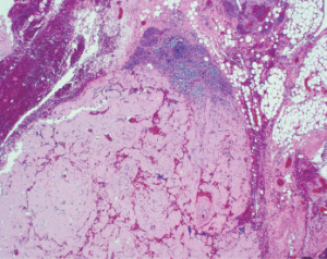
The Rheumatologist
COVID-19 News
  • Connect with us:
  • Facebook
  • Twitter
  • LinkedIn
  • YouTube
  • Feed
  • Home
  • Conditions
    • Rheumatoid Arthritis
    • SLE (Lupus)
    • Crystal Arthritis
      • Gout Resource Center
    • Spondyloarthritis
    • Osteoarthritis
    • Soft Tissue Pain
    • Scleroderma
    • Vasculitis
    • Systemic Inflammatory Syndromes
    • Guidelines
  • Resource Centers
    • Ankylosing Spondylitis Resource Center
    • Gout Resource Center
    • Rheumatoid Arthritis Resource Center
    • Systemic Lupus Erythematosus Resource Center
  • Drug Updates
    • Biologics & Biosimilars
    • DMARDs & Immunosuppressives
    • Topical Drugs
    • Analgesics
    • Safety
    • Pharma Co. News
  • Professional Topics
    • Ethics
    • Legal
    • Legislation & Advocacy
    • Career Development
      • Certification
      • Education & Training
    • Awards
    • Profiles
    • President’s Perspective
    • Rheuminations
  • Practice Management
    • Billing/Coding
    • Quality Assurance/Improvement
    • Workforce
    • Facility
    • Patient Perspective
    • Electronic Health Records
    • Apps
    • Information Technology
    • From the College
    • Multimedia
      • Audio
      • Video
  • Resources
    • Issue Archives
    • ACR Convergence
      • Systemic Lupus Erythematosus Resource Center
      • Rheumatoid Arthritis Resource Center
      • Gout Resource Center
      • Abstracts
      • Meeting Reports
      • ACR Convergence Home
    • American College of Rheumatology
    • ACR ExamRheum
    • Research Reviews
    • ACR Journals
      • Arthritis & Rheumatology
      • Arthritis Care & Research
      • ACR Open Rheumatology
    • Rheumatology Image Library
    • Treatment Guidelines
    • Rheumatology Research Foundation
    • Events
  • About Us
    • Mission/Vision
    • Meet the Authors
    • Meet the Editors
    • Contribute to The Rheumatologist
    • Subscription
    • Contact
  • Advertise
  • Search
You are here: Home / Articles / Case Report: Does a Rapid-Onset Neck Mass Have Rheumatic Origins?

Case Report: Does a Rapid-Onset Neck Mass Have Rheumatic Origins?

September 17, 2019 • By Tej Bhavsar, MD, & Nancy Joste, MD

  • Tweet
  • Email
Print-Friendly Version / Save PDF
Figure 1

Figure 1
This image shows a lymph node with near-total replacement by amorphous amyloid material (H&E, 100x).

A 54-year-old African American man arrived at the emergency department with the acute onset of a tender mass on the left side of his neck. It had been getting progressively larger for the preceding two days.

You Might Also Like
  • Case Report: An Adult-Onset Still’s Disease Mimic
  • Fellow’s Forum Case Report: Arthritis, Fever, and Weight Loss In a Teenager Suggest A Rheumatic Condition
  • Cancer-Associated Myositis: A Case Report & Review of the Literature
Explore This Issue
September 2019

History & Examination

His history included chronic right hip osteoarthritis with two surgeries performed five years prior. At his initial presentation, he noted increased fatigue the preceding few days, as well as generalized weakness and decreased appetite.

ad goes here:advert-1
ADVERTISEMENT
SCROLL TO CONTINUE

His habits included smoking a half-pack of cigarettes daily for 30 years, occasional alcohol consumption and avoidance of recreational substances. Because he had been employed by the oil industry working on scaffolding, he had traveled extensively throughout the U.S. for work, but he denied traveling outside the country or to regions where Lyme disease is known to be endemic.

On examination, he displayed a 4–5 cm non-pulsatile mass on the left side of his neck. It was moderately tender to palpation. He also had mild thyromegaly with a suggestion of thyroid nodules. His right hip was not tender and had no swelling, ecchymosis, warmth or erythema. Flexion of his hip was limited to 90º, and internal as well as external rotation was limited to approximately 15º.

ad goes here:advert-2
ADVERTISEMENT
SCROLL TO CONTINUE

Evaluation of laboratory tests revealed an elevated rheumatoid factor at 25 (upper limit of normal [ULN]: 14), and an elevated serum immunoglobulin G at 1,700 mg/dL (ULN: 1,600 mg/dL). Serum protein electrophoresis revealed a total gamma component elevation at 1.74 g/dL (ULN: 1.6 g/dL) with M-component elevation at 1.28 g/dL (ULN: 0 g/dL).

Dr. Bhavasar

Dr. Bhavasar

A computed tomography (CT) scan with contrast of the patient’s neck revealed a large, mildly heterogeneous inferior left neck and left subclavicular mass, likely reflecting a large nodal conglomerate approximately 4.5 cm anteroposterior, 7.5 cm transverse and 7.5 cm in cranio-caudal dimensions. Portions were hypoattenuating relative to muscle, perhaps reflecting cystic or necrotic areas. Calcifications seen within the mass, in conjunction with an enlarged multi­nodular thyroid gland, were most worrisome for metastatic lymphadenopathy related to thyroid cancer. An X-ray of his right hip displayed total arthroplasty without evidence of acute complication.

Fine needle aspiration of the thyroid nodule noted on the CT scan was performed and found to be consistent with a lymphocytic (Hashimoto) thyroiditis. The neck mass was biopsied, and subsequent tissue studies revealed a Gram stain with very few white blood cells, no organisms and no acid-fast bacilli. A tissue culture did not result in growth of bacteria, yeast, fungi or mycobacteria at six weeks. The biopsy demonstrated lymph nodes and fibro-adipose tissue with extensive replacement by amorphous deposits of acellular material consistent with amyloid (see Figure 1, above right). A Congo red stain was positive for polarized apple-green birefringent material, confirming the impression of amyloid (see Figure 2, p. 38). Amyloid also involved vessel walls, and areas of ossification were present. The biopsy material subsequently underwent liquid chromatography tandem mass spectroscopy at Mayo Clinic Laboratories, which subtyped the amyloid as AL (lambda) type.

ad goes here:advert-3
ADVERTISEMENT
SCROLL TO CONTINUE

Pages: 1 2 3 4 | Single Page

Filed Under: Conditions, Systemic Inflammatory Syndromes Tagged With: Amyloidosis, Cancer, case reportIssue: September 2019

You Might Also Like:
  • Case Report: An Adult-Onset Still’s Disease Mimic
  • Fellow’s Forum Case Report: Arthritis, Fever, and Weight Loss In a Teenager Suggest A Rheumatic Condition
  • Cancer-Associated Myositis: A Case Report & Review of the Literature
  • Neck Pain Patient Fact Sheet

American College of Rheumatology

Visit the official website for the American College of Rheumatology.

Visit the ACR »

ACR Convergence

Don’t miss rheumatology’s premier scientific meeting for anyone involved in research or the delivery of rheumatologic care or services.

Visit the ACR Convergence site »

Rheumatology Research Foundation

The Foundation is the largest private funding source for rheumatology research and training in the U.S.

Learn more »

The Rheumatologist newsmagazine reports on issues and trends in the management and treatment of rheumatic diseases. The Rheumatologist reaches 11,500 rheumatologists, internists, orthopedic surgeons, nurse practitioners, physician assistants, nurses, and other healthcare professionals who practice, research, or teach in the field of rheumatology.

About Us / Contact Us / Advertise / Privacy Policy / Terms of Use

  • Connect with us:
  • Facebook
  • Twitter
  • LinkedIn
  • YouTube
  • Feed

Copyright © 2006–2021 American College of Rheumatology. All rights reserved.

ISSN 1931-3268 (print)
ISSN 1931-3209 (online)

loading Cancel
Post was not sent - check your email addresses!
Email check failed, please try again
Sorry, your blog cannot share posts by email.
This site uses cookies: Find out more.