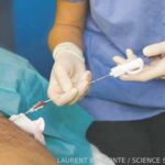
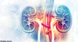
The game is changing for management & care
I met Andrea Fava, MD, when he was an intern in the medical intensive care unit. I was a year ahead of him and likely knew more about patient care—but I certainly didn’t know more about research. Nor did I have an incredible Italian accent like he did and still does. Differences aside, we became friends.
Fast forward a few years, and Andrea matched into a rheumatology fellowship at Johns Hopkins University just one year behind me. I was elated to discover that I would get to spend the next year enjoying more of that accent, as well as learning from and with him.
Fast forward another seven years, and I am overjoyed to say that Andrea and I are still friends. He is now an assistant professor of medicine, Division of Rheumatology, and director of Lupus Translational Research, Johns Hopkins University School of Medicine, Baltimore. When it comes to colleagues to call about tough systemic lupus erythematosus (SLE) cases, he tops the list.
This September for The Rheumatologist (TR), just weeks before ACR Convergence 2025, I had the chance to pick Dr. Fava’s brain about his latest research: a liquid biopsy for lupus nephritis. Sound sexy? That’s because it is.
Background
Liquid biopsy is a clever name given to a noninvasive urine proteomics test that Dr. Fava and his colleagues are developing for lupus nephritis. Put simply, urine proteomics involves the large-scale measurement of proteins in the urine, such as cytokines, growth factors, etc.1 His team is using urine proteomics to better understand and monitor lupus nephritis without relying solely on invasive renal biopsies or crude markers, such as proteinuria. This work could revolutionize the care of our patients with lupus nephritis.
Interview
TR: How does a liquid biopsy work?
Dr. Fava: Let’s start with some background. Urine is the ideal biospecimen because it’s downstream of both kidneys and collects the byproducts of kidney biology and inflammation. We’ve identified many proteins in the urine that reflect the degree of histologic activity in the kidney.2 The idea is to capture this information in a noninvasive way. We are focusing on capturing the degree of inflammation in the kidney to determine if active lupus nephritis is present.
TR: What about urine protein to creatine ratio (UPCR)? Isn’t that method good enough?


