Video: Every Case Tells a Story| Webinar: ACR/CHEST ILD Guidelines in Practice

An official publication of the ACR and the ARP serving rheumatologists and rheumatology professionals

  • Conditions
    • Axial Spondyloarthritis
    • Gout and Crystalline Arthritis
    • Myositis
    • Osteoarthritis and Bone Disorders
    • Pain Syndromes
    • Pediatric Conditions
    • Psoriatic Arthritis
    • Rheumatoid Arthritis
    • Sjögren’s Disease
    • Systemic Lupus Erythematosus
    • Systemic Sclerosis
    • Vasculitis
    • Other Rheumatic Conditions
  • FocusRheum
    • ANCA-Associated Vasculitis
    • Axial Spondyloarthritis
    • Gout
    • Psoriatic Arthritis
    • Rheumatoid Arthritis
    • Systemic Lupus Erythematosus
  • Guidance
    • Clinical Criteria/Guidelines
    • Ethics
    • Legal Updates
    • Legislation & Advocacy
    • Meeting Reports
      • ACR Convergence
      • Other ACR meetings
      • EULAR/Other
    • Research Rheum
  • Drug Updates
    • Analgesics
    • Biologics/DMARDs
  • Practice Support
    • Billing/Coding
    • EMRs
    • Facility
    • Insurance
    • QA/QI
    • Technology
    • Workforce
  • Opinion
    • Patient Perspective
    • Profiles
    • Rheuminations
      • Video
    • Speak Out Rheum
  • Career
    • ACR ExamRheum
    • Awards
    • Career Development
  • ACR
    • ACR Home
    • ACR Convergence
    • ACR Guidelines
    • Journals
      • ACR Open Rheumatology
      • Arthritis & Rheumatology
      • Arthritis Care & Research
    • From the College
    • Events/CME
    • President’s Perspective
  • Search

ACR Image Competition 2021 Results, Part 7

Kurt Blake, MBBS  |  Issue: September 2022  |  September 6, 2022

The featured image from North America. (Click to enlarge.)

Saddle Nose & Cauliflower Ear Deformities in Relapsing Polychondritis

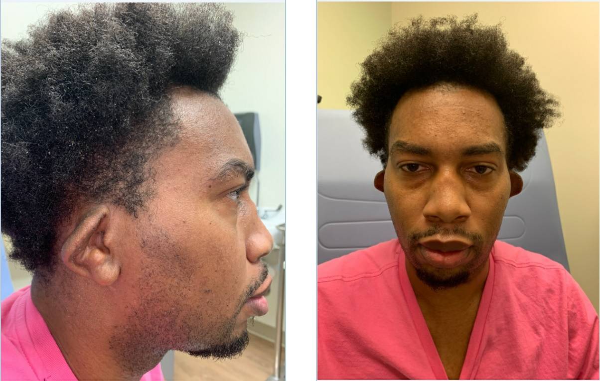
These images depict a 32-year-old man who presented with five weeks of left-sided hearing loss, weight loss and discomfort in the nose, ear, chest wall and knee. He had an erythrocyte sedimentation rate (ESR) of 120 mm/hr, and a C-reactive protein level of 225.4 mg/L. The photographs show his nose and ear deformities.


Born and raised in Jamaica, Kurt Blake, MBBS, finished medical school at the University of the West Indies and worked with an orthopedic team in a rural hospital. Later, he joined the faculty of St. George’s University, Grenada, West Indies. He is now pursuing a fellowship in rheumatology at the University of Alabama at Birmingham.

ad goes here:advert-1
ADVERTISEMENT
SCROLL TO CONTINUE

About the Contest

The Rheumatology Image Library is a highly accessed teaching resource. However, images showing manifestations of rheumatic disease on skin of color are underrepresented, creating a significant educational gap. The 2021 Image Competition, held in conjunction with ACR Convergence 2021, encouraged the global rheumatology community to submit images that will help healthcare providers identify rheumatic disease manifestations in skin of color. Here, we depict the image featured from North America In coming issues, we’ll publish the winning images from other regions. The images judged Best Overall and People’s Choice, and others, appeared in previous issues.

Share: 

Filed under:ConditionsProfessional Topics Tagged with:biascauliflower eardiagnostic imagingDiversityhealthcare disparitiesImage CompetitionRacismrelapsing polychondritissaddle nose deformity

Related Articles

    ACR Image Competition 2021 Results, Part 5

    July 13, 2022

    Rheumatic Diseases of Childhood: Juvenile Dermatomyositis with Calcinosis Cutis These images depict a 14-year-old boy with a two-year history of proximal muscle weakness affecting both upper and lower limbs, and a skin rash affecting his face. He was diagnosed with juvenile dermatomyositis and developed calcinosis over both legs with skin infection and ulceration. Plain X-ray…

    ACR Image Competition 2021 Results, Part 6

    August 14, 2022

    Featured Image from Europe & Central Asia Rheumatic Diseases of Childhood: Scleroderma These images depict a patient with a clinical presentation of juvenile dermatomyositis with typical skin manifestations of Gottron’s papules over the extensor surfaces of the joints, as well as hyperpigmentation. ad goes here:advert-1ADVERTISEMENTSCROLL TO CONTINUE Yulia Vyzhga, MD, PhD, is a pediatric rheumatologist…

    ACR Image Competition 2021 Results, Part 4

    June 14, 2022

    Lichenoid Cutaneous Lupus Erythematosus Mimicking Acanthosis Nigricans The photos depict a 45-year-old woman who presented to the Lupus Clinic of the University of São Paulo, Brazil, with lesions closely resembling acanthosis nigricans on her neck (A and B). The lesions had been present for four months. The patient had lived with systemic lupus erythematosus (SLE)…

    Images as Teaching Tools: A Call for Entries in 2021 ACR Image Competition

    June 8, 2021

    As part of the ACR’s pledge to be a leader for inclusion and change for members, trainees, staff and rheumatology patients, the ACR is holding a special image competition in conjunction with ACR Convergence 2021 dedicated exclusively to images of rheumatic disease in skin of color. “We started discussing the lack of diversity in our…

  • About Us
  • Meet the Editors
  • Issue Archives
  • Contribute
  • Advertise
  • Contact Us
  • Copyright © 2025 by John Wiley & Sons, Inc. All rights reserved, including rights for text and data mining and training of artificial technologies or similar technologies. ISSN 1931-3268 (print). ISSN 1931-3209 (online).
  • DEI Statement
  • Privacy Policy
  • Terms of Use
  • Cookie Preferences