Video: Every Case Tells a Story| Webinar: ACR/CHEST ILD Guidelines in Practice

An official publication of the ACR and the ARP serving rheumatologists and rheumatology professionals

  • Conditions
    • Axial Spondyloarthritis
    • Gout and Crystalline Arthritis
    • Myositis
    • Osteoarthritis and Bone Disorders
    • Pain Syndromes
    • Pediatric Conditions
    • Psoriatic Arthritis
    • Rheumatoid Arthritis
    • Sjögren’s Disease
    • Systemic Lupus Erythematosus
    • Systemic Sclerosis
    • Vasculitis
    • Other Rheumatic Conditions
  • FocusRheum
    • ANCA-Associated Vasculitis
    • Axial Spondyloarthritis
    • Gout
    • Psoriatic Arthritis
    • Rheumatoid Arthritis
    • Systemic Lupus Erythematosus
  • Guidance
    • Clinical Criteria/Guidelines
    • Ethics
    • Legal Updates
    • Legislation & Advocacy
    • Meeting Reports
      • ACR Convergence
      • Other ACR meetings
      • EULAR/Other
    • Research Rheum
  • Drug Updates
    • Analgesics
    • Biologics/DMARDs
  • Practice Support
    • Billing/Coding
    • EMRs
    • Facility
    • Insurance
    • QA/QI
    • Technology
    • Workforce
  • Opinion
    • Patient Perspective
    • Profiles
    • Rheuminations
      • Video
    • Speak Out Rheum
  • Career
    • ACR ExamRheum
    • Awards
    • Career Development
  • ACR
    • ACR Home
    • ACR Convergence
    • ACR Guidelines
    • Journals
      • ACR Open Rheumatology
      • Arthritis & Rheumatology
      • Arthritis Care & Research
    • From the College
    • Events/CME
    • President’s Perspective
  • Search

Case Report: A Rare But Severe Complication of Dermatomyositis

Akrithi Udupa, MD, Paul McIntosh, MD, Thomas J. Cummings, MD, & Lisa Criscione-Schreiber, MD, Med  |  Issue: May 2021  |  May 13, 2021

Dermatomyositis is an uncommon autoimmune condition involving skeletal muscle characterized by subacute onset of progressive weakness, intramuscular inflammatory infiltrates and the presence of myositis-specific autoantibodies.1 Immune-mediated myopathies may exert some pathogenic effects on the muscle tissue by targeting the microvasculature.1 Capillary inflammation, fragility and loss may contribute to heightened bleeding events in these patients.

Here, we discuss a case of acute hemorrhagic dermatomyositis, a rare, highly morbid complication of dermatomyositis.

ad goes here:advert-1
ADVERTISEMENT
SCROLL TO CONTINUE

Case History

A previously healthy, 66-year-old man presented with gradual onset of painfully swollen and stiff upper and lower extremities, diffuse rash and progressive weakness. He worked as a custodian and reported that he’d had difficulty lifting trash cans and climbing stairs for the past two months. He also reported that during the preceding two weeks he’d been struggling to reach for his seatbelt and had difficulty getting in and out of his truck. He ultimately presented for medical attention because he could no longer ambulate independently.

Over the past year, he had experienced fatigue, episodic dizziness, daytime and nighttime sweats, reduced appetite, trouble swallowing and unintentional weight loss. He lived alone and had not seen a medical provider for more than five decades until the previous month, following an emergency department visit. He had a 45-pack-year smoking history and a remote history of heavy alcohol use.

ad goes here:advert-2
ADVERTISEMENT
SCROLL TO CONTINUE

Vital signs in the emergency department were normal. An examination revealed a chronically ill-appearing man in no acute distress. He had multiple, erythematous, pearly nodules up to four centimeters in diameter on his left forehead, right eyebrow and left neck, suggestive of basal cell carcinoma (see Figure 1).

An erythematous maculopapular poikilodermic rash was present on his scalp, forehead, cheeks, neck, chest, upper back, upper arms, and dorsal aspects of his metacarpophalangeal and interphalangeal joints of his digits. He had painful periungual erythema of his fingernails.

His neurologic exam was notable for symmetric proximal weakness, with his upper extremities being weaker than his lower extremities. Manual muscle testing revealed the following Medical Research Council (MRC) grades: cervical flexion and extension 5/5, deltoids 2/5, biceps and triceps 3/5, wrist extensors and flexors 5/5 apart from 4+/5 left wrist extension, finger abduction 4+/5, finger flexion 5/5, hip flexion 3/5, quadriceps 5-/5, hamstrings 4+/5, anterior tibialis 5/5, gastrocnemius 5/5.

The physical examination revealed multiple, large, erythematous pearly nodules suggestive of basal cell carcinoma, as shown on his left temple, and a diffuse poikilodermic rash (maroon arrows).

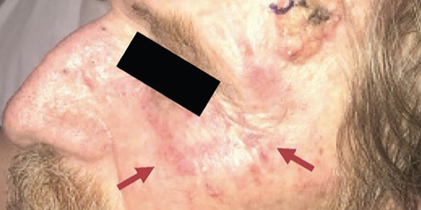
Figure 1. The physical examination revealed multiple, large, erythematous pearly nodules suggestive of basal cell carcinoma, as shown on his left temple, and a diffuse poikilodermic rash (maroon arrows).

He had no ocular, facial or bulbar weakness or sensory deficits. His deep tendon reflexes were trace at the biceps, brachioradialis and Achilles tendons. His patellar reflexes were 3+ with crossed adduction in both limbs. The remainder of the exam was unremarkable.

Emergency department laboratory testing from four weeks before was notable for normal renal function, normal blood counts, aspartate aminotransferase (AST) 313 IU/L (normal: 10–50 IU/L) and alanine aminotransferase (ALT) 124 IU/L (normal: 10–44 IU/L).

At follow-up with his primary care provider four days later, his liver transaminases were persistently elevated: AST 301 IU/L, ALT 134 IU/L and alkaline phosphatase (ALP) 116 U/L (normal: 40–129 U/L). Other abnormally elevated laboratory studies at this visit included C-reactive protein (CRP) 30 mg/L, anti-nuclear antibody (ANA) 1:320 with speckled pattern, anti-smooth muscle antibody (ASMA) 54 U, anti-mitochondrial antibody (AMA) 43.8 U, rheumatoid factor (RF) 57.8 IU/mL. Lyme IgG/IgM and Rocky Mountain Spotted Fever IgM screening tests fell within equivocal range.

Page: 1 2 3 4 5 6 | Single Page
Share: 

Filed under:ConditionsMyositis Tagged with:case reportdermatomyositis (DM)hemorrhagic dermatomyositis

Related Articles

    New Tools for Myositis Diagnosis, Classification & Management

    April 15, 2019

    CHICAGO—At Hot Topics in Myositis, a session at the 2018 ACR/ARHP Annual Meeting, three experts discussed new classification criteria for idiopathic inflammatory myopathies (IIM) and offered practical primers on overlap myositis conditions and inclusion body myositis (IBM). New Myositis Classification Criteria After a 10-year development process, the new EULAR/ACR Classification Criteria for Adult and Juvenile…

    Autoantibodies in Autoimmune Myopathy

    Autoantibodies in Autoimmune Myopathy

    September 18, 2017

    In recent years, scientists and clinicians have learned a great deal about autoantibodies occurring in idiopathic inflammatory myopathies (IIMs). These new discoveries have reshaped our understanding of distinct clinical pheno­types in IIMs. Scientists continue to learn more about how these auto­antibodies shape pathophysiology, diagnosis, disease monitoring, prognosis and optimum treatment. Moving forward, these autoantibodies will…

    Tashatuvango / shutterstock.com

    Myositis-Specific Antibodies Identified

    January 16, 2020

    The idiopathic inflammatory myopathies (IIM) encompass eight categories: 1) dermatomyositis (DM) in adults, 2) juvenile dermatomyositis, 3) amyopathic DM, 4) cancer-associated DM, 5) polymyositis, 6) immune-mediated necrotizing myopathy, 7) inclusion body myositis, and 8) overlap myositis.1 These categories help classify the myopathies based on clinical and histologic features. The incidence of IIM is estimated at…

    Myositis Mysteries

    January 1, 2008

    Why isn’t my myositis patient getting better?

  • About Us
  • Meet the Editors
  • Issue Archives
  • Contribute
  • Advertise
  • Contact Us
  • Copyright © 2025 by John Wiley & Sons, Inc. All rights reserved, including rights for text and data mining and training of artificial technologies or similar technologies. ISSN 1931-3268 (print). ISSN 1931-3209 (online).
  • DEI Statement
  • Privacy Policy
  • Terms of Use
  • Cookie Preferences