Video: Every Case Tells a Story| Webinar: ACR/CHEST ILD Guidelines in Practice

An official publication of the ACR and the ARP serving rheumatologists and rheumatology professionals

  • Conditions
    • Axial Spondyloarthritis
    • Gout and Crystalline Arthritis
    • Myositis
    • Osteoarthritis and Bone Disorders
    • Pain Syndromes
    • Pediatric Conditions
    • Psoriatic Arthritis
    • Rheumatoid Arthritis
    • Sjögren’s Disease
    • Systemic Lupus Erythematosus
    • Systemic Sclerosis
    • Vasculitis
    • Other Rheumatic Conditions
  • FocusRheum
    • ANCA-Associated Vasculitis
    • Axial Spondyloarthritis
    • Gout
    • Psoriatic Arthritis
    • Rheumatoid Arthritis
    • Systemic Lupus Erythematosus
  • Guidance
    • Clinical Criteria/Guidelines
    • Ethics
    • Legal Updates
    • Legislation & Advocacy
    • Meeting Reports
      • ACR Convergence
      • Other ACR meetings
      • EULAR/Other
    • Research Rheum
  • Drug Updates
    • Analgesics
    • Biologics/DMARDs
  • Practice Support
    • Billing/Coding
    • EMRs
    • Facility
    • Insurance
    • QA/QI
    • Technology
    • Workforce
  • Opinion
    • Patient Perspective
    • Profiles
    • Rheuminations
      • Video
    • Speak Out Rheum
  • Career
    • ACR ExamRheum
    • Awards
    • Career Development
  • ACR
    • ACR Home
    • ACR Convergence
    • ACR Guidelines
    • Journals
      • ACR Open Rheumatology
      • Arthritis & Rheumatology
      • Arthritis Care & Research
    • From the College
    • Events/CME
    • President’s Perspective
  • Search

Case Report: Diaphragm Ultrasound Reveals Shrinking Lung Syndrome

Mery Deeb, MD, Taro Minami, MD, Michael Stanchina, MD, Elias Jabbour, MD, & Jan Karczewski, MD  |  Issue: July 2024  |  July 9, 2024

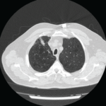
The patient was referred to a pulmonary specialist, who performed diaphragmatic ultrasound. The ultrasound revealed both reduction in diaphragm dome excursion and in diaphragm thickening fraction, consistent with bilateral diaphragm dysfunction, confirming diaphragmatic dysfunction and a diagnosis of shrinking lung syndrome (see Figure 1). Follow-up high-resolution CT showed no evidence of interstitial lung disease. Mycophenolate mofetil was initiated, in addition to prednisone.

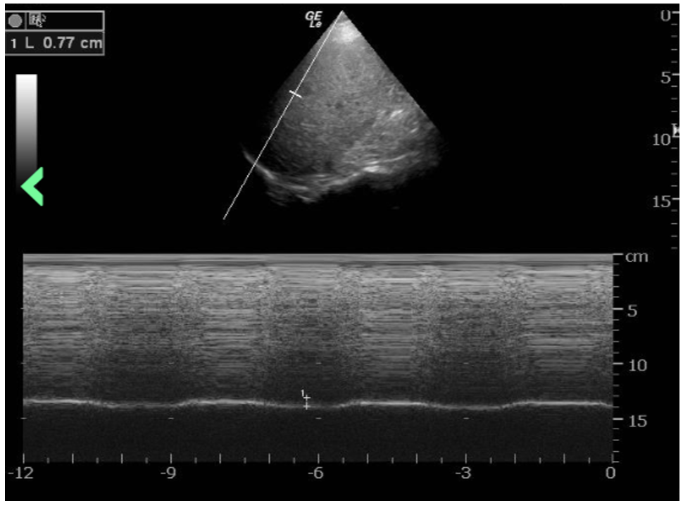
Figure 1: The ultrasound revealed a reduction in diaphragm dome excursion of 0.77 cm (normal value for deep inspiration: 4.8 cm) and in diaphragm thickening fraction, consistent with bilateral diaphragm dysfunction. (Click to enlarge.)

Repeat PFTs three months later demonstrated improvement, with FVC post 39%, FEV1 post 37%, FEV1/FVC post 92% predicted, TLC 50%, DLCO 38%, reduction in mean inspiratory pressure and decline of vital capacity from a seated to supine position.

ad goes here:advert-1
ADVERTISEMENT
SCROLL TO CONTINUE

Discussion

Diagnosis of shrinking lung syndrome usually follows the diagnosis of SLE, with a mean interval of 66.7 months.2 This case is atypical because shrinking lung syndrome was part of the initial presentation of systemic lupus erythematosus. This added to diagnostic uncertainty in this case.

It is important to highlight the role of ultrasonography of the diaphragm in our case, which confirmed diaphragm involvement and assisted us in making the diagnosis of shrinking lung syndrome. Upon our patient’s initial presentation to the hospital, there was concern for a possible overlying acute process, such as neumonia. Although additional CT scans showed resolution of parenchymal lung abnormalities, the patient continued to have severe dyspnea. Subsequent PFTs showed restrictive lung defect and resulted in an unclear clinical picture.

ad goes here:advert-2
ADVERTISEMENT
SCROLL TO CONTINUE

Fortunately, ultrasonography is a noninvasive and rapid bedside test that can provide real-time data on diaphragmatic function without subjecting patients to radiation. It has a sensitivity and specificity of more than 90% for diagnosis of diaphragmatic dysfunction.3-5 Real-time ultrasound in this case confirmed diaphragmatic dysfunction and enabled immediate initiation of immunosuppressive therapy, rather than waiting for repeat chest imaging.

Although the exact pathogenesis of shrinking lung syndrome is unclear, several hypotheses have been proposed, including that it may result from surfactant deficiency, isolated diaphragmatic myopathy, phrenic neuropathy, chest wall dysfunction or pleural adhesions.6

Recent data suggest that shrinking lung syndrome may be a complication of pleural inflammation. Inflammation, particularly around the regions of the diaphragmatic pleura or a pleural region near the diaphragm, could lead to chronic lung hypoinflation and loss of compliance due to activation of local neuroreflexes, which inhibit diaphragm function and eventually cause diaphragmatic remodeling.7

Page: 1 2 3 4 5 | Single Page
Share: 

Filed under:ConditionsSystemic Lupus Erythematosus Tagged with:case reportdyspneashrinking lung syndromeSLE Resource CenterUltrasound

Related Articles
    Medium-sized blood vessels are seen in the dermis and at the dermal-subcutaneous junction. The black arrow identifies a venule, and the blue arrow points to a fibrin deposition in the vessel wall. Extravasated neutrophils and erythrocytes are seen in the interstitium.

    Case Report: A Polyarteritis Nodosa Conundrum

    March 14, 2022

    Polyarteritis nodosa (PAN) is a systemic necrotizing vasculitis that typically affects medium-sized muscular arteries. The clinical subsets of PAN are idiopathic, generalized, secondary hepatitis B virus (HBV) associated and cutaneous PAN. These clinical subsets are important because of their therapeutic implications. Virtually any organ system can be affected in generalized PAN, but this vasculitis tends…

    Case Report: Perplexing Pulmonary Nodules

    March 14, 2022

    Pulmonary nodules are com­mon; most are benign, but the differential diagnosis is broad and includes life-threatening possibilities.1 Our patient is a former smoker who has a history of a complex autoimmune disease and multiple pulmonary nodules. This case was challenging, but clinical, radiographic and histologic clues helped lead to the correct diagnosis. Case Presentation The…

    APS: What Rheumatologists Should Know about Hughes Syndrome

    February 17, 2016

    The problem that dogs the work of all of those treating patients with antiphospholipid syndrome (APS) is the apparent lack of knowledge of the syndrome, both by the general public, as well as by swaths of the medical fraternity. Perhaps it was ever thus—a syndrome less than 40 years old could be described as new,…

    Case Report: Interstitial Lung Disease with Positive ANCA Test

    October 13, 2015

    “Worst of all is the pain in my calves,” she said. “It feels like burning deep inside.” So began my first encounter with a 69-year-old woman who was referred to rheumatology clinic for evaluation of two months of constitutional symptoms and a positive ANCA test, which had been ordered by her primary care doctor. Her…

  • About Us
  • Meet the Editors
  • Issue Archives
  • Contribute
  • Advertise
  • Contact Us
  • Copyright © 2025 by John Wiley & Sons, Inc. All rights reserved, including rights for text and data mining and training of artificial technologies or similar technologies. ISSN 1931-3268 (print). ISSN 1931-3209 (online).
  • DEI Statement
  • Privacy Policy
  • Terms of Use
  • Cookie Preferences