Video: Every Case Tells a Story| Webinar: ACR/CHEST ILD Guidelines in Practice

An official publication of the ACR and the ARP serving rheumatologists and rheumatology professionals

  • Conditions
    • Axial Spondyloarthritis
    • Gout and Crystalline Arthritis
    • Myositis
    • Osteoarthritis and Bone Disorders
    • Pain Syndromes
    • Pediatric Conditions
    • Psoriatic Arthritis
    • Rheumatoid Arthritis
    • Sjögren’s Disease
    • Systemic Lupus Erythematosus
    • Systemic Sclerosis
    • Vasculitis
    • Other Rheumatic Conditions
  • FocusRheum
    • ANCA-Associated Vasculitis
    • Axial Spondyloarthritis
    • Gout
    • Psoriatic Arthritis
    • Rheumatoid Arthritis
    • Systemic Lupus Erythematosus
  • Guidance
    • Clinical Criteria/Guidelines
    • Ethics
    • Legal Updates
    • Legislation & Advocacy
    • Meeting Reports
      • ACR Convergence
      • Other ACR meetings
      • EULAR/Other
    • Research Rheum
  • Drug Updates
    • Analgesics
    • Biologics/DMARDs
  • Practice Support
    • Billing/Coding
    • EMRs
    • Facility
    • Insurance
    • QA/QI
    • Technology
    • Workforce
  • Opinion
    • Patient Perspective
    • Profiles
    • Rheuminations
      • Video
    • Speak Out Rheum
  • Career
    • ACR ExamRheum
    • Awards
    • Career Development
  • ACR
    • ACR Home
    • ACR Convergence
    • ACR Guidelines
    • Journals
      • ACR Open Rheumatology
      • Arthritis & Rheumatology
      • Arthritis Care & Research
    • From the College
    • Events/CME
    • President’s Perspective
  • Search

Case Report: Lymphocytic Vasculitis of the Central Nervous System

Gbemisola Olayemi, MD, Evangeline Scopelitis, MD, & Jerald M. Zakem, MD  |  Issue: January 2019  |  January 17, 2019

Vasculitis is a group of chronic inflammatory diseases in which the blood vessel is the target of an immune reaction. They can be secondary to connective tissue disease, idiopathic or due to infection, neoplasm or drugs.1 Primary angiitis of the central nervous system (PACNS) is a rare syndrome characterized by inflammatory cell infiltration and necrosis of small- and medium-sized blood vessels of the brain parenchyma, spinal cord and leptomeninges.2 It was reported in 1922 by Harbitz as an unusual type of arteritis associated with multinucleate giant cell arteritis of the small meninges and cerebrum and was first reported as a distinct clinical-pathologic entity in an article by Cravioto and Feigin in 1959.3,4

The estimated annual incidence rate of PACNS is 2.4 cases per 1 million person-years. Middle-aged men are most often affected by PACNS, with a median age at onset of approximately 50 years and a male-to-female ratio of approximately 2:1.5 We describe the clinical, imaging and histopathological findings in a patient with PACNS.

ad goes here:advert-1
ADVERTISEMENT
SCROLL TO CONTINUE

Case Presentation

A 37-year-old woman presented at the emergency department with altered mental status, bilateral vision loss and weakness. She had a past medical history significant for multiple sclerosis on interferon beta, type 1 diabetes, hypertension, stage 4 chronic kidney disease and vitamin D deficiency. The patient was in her usual state of health on the morning of presentation.

Around 7:30 a.m. she held a phone conversation with her father who had traveled to her home to transport her to a previously scheduled doctor’s appointment. She did not answer the door, but her father could hear her fumbling with the lock. He contacted the property manager and gained access to the home at approximately 8:45 a.m. At that time, he found her standing at the doorway, confused. When he questioned his daughter, she replied in streams of unintelligible, mumbled speech. Her father helped her to a couch and noted she had a poorly coordinated gait.

ad goes here:advert-2
ADVERTISEMENT
SCROLL TO CONTINUE

Based on her symptoms, her father took her to the emergency department for evaluation. The patient had no recollection of the morning’s events and reported regaining her orientation and awareness on the way to the hospital.

On arrival at the emergency department, she was afebrile and tachycardic (HR 100 beats per minute), with a blood pressure of 125/75 mmHG. She was breathing normally, with oxygen saturation of 99% on room air.

When the patient was admitted to the medical ward, her vision loss had resolved. The physical exam was notable for normal mental status, right upper extremity mild dysmetria, with 4/5 strength in the right upper extremity and right lower extremity, with normal sensation and tone. There was no evidence of meningitis or focal neurologic signs.

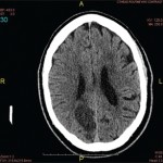
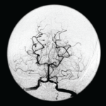
Imaging was obtained and included a computed tomography (CT) of her head, which showed several lesions suspicious for acute or subacute infarction.

Magnetic resonance imaging (MRI) of her brain showed widespread foci of restricted diffusion with corresponding low apparent diffusion coefficient (ADC) signal throughout both cerebral and cerebellar hemispheres, most suggestive of acute infarcts. The distribution was atypical for multiple sclerosis, so they were not thought to be demyelinating plaques, despite her past history. Scattered T2/FLAIR signal hyperintensities noted within the left cerebellum and right cerebral peduncle were suggestive of remote infarcts (see Figures 1 and 2).

Page: 1 2 3 4 5 6 7 | Single Page
Share: 

Filed under:ConditionsVasculitis Tagged with:central nervous system vasculitiscyclophosphamideMethylprednisolonePrimary Angiitis of the Central Nervous System

Related Articles

    Meet the Challenge of Primary CNS Vasculitis

    September 1, 2011

    Diagnosis and treatment of this rare and poorly understood condition

    Rheumatology Case Report: When Moyamoya Disease Mimicks Primary Central Nervous System Vasculitis

    November 16, 2015

    Case report: A 60-year-old Hispanic male with poorly controlled hypertension was sent from the primary care clinic for evaluation of malignant hypertension with a systolic blood pressure above 200 mmHg. His symptoms at the time of presentation included episodic confusion, worsening vision and an unsteady gait. A head computed tomography (CT) scan showed a subacute…

    7 Key Insights Into the Evaluation of Central Nervous System Vasculitis

    May 13, 2021

    Many a rheumatology consult has centered on a perplexing question: Does this patient have central nervous system (CNS) vasculitis? At the 2021 ACR State-of-the-Art Clinical Symposium, Rula Hajj-Ali, MD, FACP, professor of medicine and associate director of vasculitis care and research, Cleveland Clinic Lerner College of Medicine, discussed this topic in detail, providing a series…

    2014 ACR/ARHP Annual Meeting: Diagnosing PACNS and Its Mimics

    February 1, 2015

    Accurate diagnosis for primary angiitis of the central nervous system requires close scrutiny, team effort

  • About Us
  • Meet the Editors
  • Issue Archives
  • Contribute
  • Advertise
  • Contact Us
  • Copyright © 2025 by John Wiley & Sons, Inc. All rights reserved, including rights for text and data mining and training of artificial technologies or similar technologies. ISSN 1931-3268 (print). ISSN 1931-3209 (online).
  • DEI Statement
  • Privacy Policy
  • Terms of Use
  • Cookie Preferences