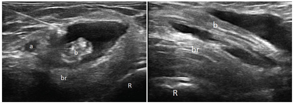
Figure 1 (left): Transverse 18 MHz ultrasound image over the right antecubital fossa at a level distal to the joint line. The left side of the image is medial, and the right side is lateral (R: radius, br: brachialis muscle, a: brachial artery, b: biceps tendon). & Figure 2 (above): Longitudinal 18 MHz ultrasound image over the right antecubital fossa at a level distal to the joint line. The left side of the image is proximal; the right side is distal (R: radius, br: brachialis muscle, b: biceps tendon).