Video: Every Case Tells a Story| Webinar: ACR/CHEST ILD Guidelines in Practice

An official publication of the ACR and the ARP serving rheumatologists and rheumatology professionals

  • Conditions
    • Axial Spondyloarthritis
    • Gout and Crystalline Arthritis
    • Myositis
    • Osteoarthritis and Bone Disorders
    • Pain Syndromes
    • Pediatric Conditions
    • Psoriatic Arthritis
    • Rheumatoid Arthritis
    • Sjögren’s Disease
    • Systemic Lupus Erythematosus
    • Systemic Sclerosis
    • Vasculitis
    • Other Rheumatic Conditions
  • FocusRheum
    • ANCA-Associated Vasculitis
    • Axial Spondyloarthritis
    • Gout
    • Psoriatic Arthritis
    • Rheumatoid Arthritis
    • Systemic Lupus Erythematosus
  • Guidance
    • Clinical Criteria/Guidelines
    • Ethics
    • Legal Updates
    • Legislation & Advocacy
    • Meeting Reports
      • ACR Convergence
      • Other ACR meetings
      • EULAR/Other
    • Research Rheum
  • Drug Updates
    • Analgesics
    • Biologics/DMARDs
  • Practice Support
    • Billing/Coding
    • EMRs
    • Facility
    • Insurance
    • QA/QI
    • Technology
    • Workforce
  • Opinion
    • Patient Perspective
    • Profiles
    • Rheuminations
      • Video
    • Speak Out Rheum
  • Career
    • ACR ExamRheum
    • Awards
    • Career Development
  • ACR
    • ACR Home
    • ACR Convergence
    • ACR Guidelines
    • Journals
      • ACR Open Rheumatology
      • Arthritis & Rheumatology
      • Arthritis Care & Research
    • From the College
    • Events/CME
    • President’s Perspective
  • Search

Did Shiitake Mushrooms Induce Immune-Mediated Necrotizing Myopathy?

Takeshi Yoshida, MD, Hiroaki Chikazawa, MD, Yoshitaka Kumon, MD, PhD, & Ichizo Nishino, MD, PhD  |  Issue: April 2019  |  April 15, 2019

A: HE staining. One necrotic fiber was observed (large arrow). Some muscle fibers with internal nuclei were observed (small arrow). B: ATPase staining. A few type 2C fibers were observed (arrows). C: MHC Class I staining. Moderate upregulation of sarcolemmal MHC class I molecule was seen.

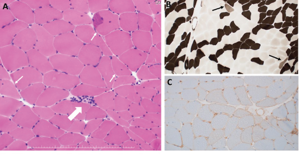
Figure 1: Histopathological Findings of Left Biceps Brachii
A: HE staining. One necrotic fiber was observed (large arrow). Some muscle fibers with internal nuclei were observed (small arrow). B: ATPase staining. A few type 2C fibers were observed (arrows). C: MHC Class I staining. Moderate upregulation of sarcolemmal MHC class I molecule was seen.

A diagnosis of immune-mediated necrotizing myopathy associated with these two antibodies was made. We started 50 mg a day of prednisolone orally, as well as cessation of mushroom intake. His symptoms and CK levels quickly improved. We tapered the prednisolone and started azathioprine. Although we discontinued azathioprine because of side effects, his symptoms remained stable. Eighteen months later, we successfully discontinued prednisolone and are now observing his symptoms without treatment.

In East Asia, the rate of statin-naive, anti-HMGCR antibody-positive immune-mediated necrotizing myopathy is much higher than in North America, raising a possibility that different triggering materials may be associated with immune-mediated necrotizing myopathy.

Discussion

Statins are believed to trigger auto­antibody production against HMGCR in individuals who have a susceptibility.8 Regenerating fibers are known to overexpress HMGCR. Therefore, statin-induced myofiber necrosis and generation may well provide more antigens to produce pathogenic autoantibodies, creating a feed-forward loop.2,7 The binding of statins to HMGCR may also change the conformation of the protein, leading to the generation of new epitopes to which the immune system is not tolerant.2

In East Asia, the rate of statin-naive, anti-HMGCR antibody-positive immune-mediated necrotizing myopathy is much higher than in North America, raising a possibility that different triggering materials may be associated with immune-mediated necrotizing myopathy—especially in patients in East Asia.3-5 In this regard, it may be an attractive hypothesis that the foods containing natural HMGCR inhibitors, such as shiitake mushrooms, red yeast rice and pu’erh tea (a variety of fermented tea), which are apparently consumed more frequently in the region, trigger the disease in this statin-naive population. Further, infantile-onset immune-mediated necrotizing myopathy associated with anti-HMGCR autoantibodies has been found in patients from Japan.6 This is interesting considering shiitake mushrooms are often used as a weaning food in Japan.

ad goes here:advert-2
ADVERTISEMENT
SCROLL TO CONTINUE

Although shiitake mushrooms have been shown to have an inhibitory effect on HMGCR, the specific molecule responsible for this effect has not been found. Gil-Ramirez, et al. showed that among water extracts obtained from 26 different mushroom species, Lentinula edodes was one of the strongest in exerting HMGCR inhibitory activity.9 The same authors also reported that the administration of extracts from Lentinula edodes to mice modulated the transcriptional profile of genes involved in the cholesterol metabolism, similarly to simvastatin, indicating shiitake mushrooms exert inhibitory activity on HMGCR similar to that of statins and may well trigger autoimmunity to HMGCR just as statins do.10

In the case presented here, simultaneous cessation of mushroom ingestion and treatment with glucocorticoids and an immunosuppressive agent led to complete remission of disease activity. Successful treatment of anti-HMGCR antibody-positive immune-mediated necrotizing myopathy usually requires not only cessation of the offending agent but also treatment with immunosuppressive agents, just as in this case.2,10

Our patient showed the presence of both of anti-HMGCR and anti-SRP antibodies. Although these two autoantibodies are considered to be mutually exclusive, the ELISA system we used has very high specificity in detecting both of them.11 In a large case series in Japan, among 387 patients with idiopathic inflammatory myopathies other than inclusion body myositis, the frequencies of anti-SRP and anti-HMGCR antibodies were 18% and 12%, respectively, and only one patient had both autoantibodies. Details of this case were not discussed in the literature.4 We need to wait for more reports of similar cases to know the exact pathomechanism of co-existence of these two antibodies.

ad goes here:advert-1
ADVERTISEMENT
SCROLL TO CONTINUE

Page: 1 2 3 4 | Single Page
Share: 

Filed under:ConditionsOther Rheumatic Conditions Tagged with:2018 ACR/ARHP Annual Meetingcase reportDietmushroomsnecrotizing myopathyshiitake mushrooms

Related Articles

    Fellows’ Forum Case Report: Necrotizing Autoimmune Myopathy

    December 18, 2017

    Necrotizing autoimmune myopathy (NAM) is a relatively recently discovered subgroup of inflammatory myopathies. NAM is characterized by predominant muscle fiber necrosis and regeneration with little or no inflammation.1 One subgroup of NAM is 3-hydroxy-3-methylglutaryl-CoA reductase antibody (HMGCR Ab)-related immune-mediated necrotizing myopathy (IMNM), which occurs (rarely) after statin exposure, with a rough incidence of two per…

    New Tools for Myositis Diagnosis, Classification & Management

    April 15, 2019

    CHICAGO—At Hot Topics in Myositis, a session at the 2018 ACR/ARHP Annual Meeting, three experts discussed new classification criteria for idiopathic inflammatory myopathies (IIM) and offered practical primers on overlap myositis conditions and inclusion body myositis (IBM). New Myositis Classification Criteria After a 10-year development process, the new EULAR/ACR Classification Criteria for Adult and Juvenile…

    Drug Updates

    January 1, 2009

    Information on safety, labeling changes, and pharmaceutical research

    Autoantibodies in Autoimmune Myopathy

    Autoantibodies in Autoimmune Myopathy

    September 18, 2017

    In recent years, scientists and clinicians have learned a great deal about autoantibodies occurring in idiopathic inflammatory myopathies (IIMs). These new discoveries have reshaped our understanding of distinct clinical pheno­types in IIMs. Scientists continue to learn more about how these auto­antibodies shape pathophysiology, diagnosis, disease monitoring, prognosis and optimum treatment. Moving forward, these autoantibodies will…

  • About Us
  • Meet the Editors
  • Issue Archives
  • Contribute
  • Advertise
  • Contact Us
  • Copyright © 2025 by John Wiley & Sons, Inc. All rights reserved, including rights for text and data mining and training of artificial technologies or similar technologies. ISSN 1931-3268 (print). ISSN 1931-3209 (online).
  • DEI Statement
  • Privacy Policy
  • Terms of Use
  • Cookie Preferences