Video: Every Case Tells a Story| Webinar: ACR/CHEST ILD Guidelines in Practice

An official publication of the ACR and the ARP serving rheumatologists and rheumatology professionals

  • Conditions
    • Axial Spondyloarthritis
    • Gout and Crystalline Arthritis
    • Myositis
    • Osteoarthritis and Bone Disorders
    • Pain Syndromes
    • Pediatric Conditions
    • Psoriatic Arthritis
    • Rheumatoid Arthritis
    • Sjögren’s Disease
    • Systemic Lupus Erythematosus
    • Systemic Sclerosis
    • Vasculitis
    • Other Rheumatic Conditions
  • FocusRheum
    • ANCA-Associated Vasculitis
    • Axial Spondyloarthritis
    • Gout
    • Psoriatic Arthritis
    • Rheumatoid Arthritis
    • Systemic Lupus Erythematosus
  • Guidance
    • Clinical Criteria/Guidelines
    • Ethics
    • Legal Updates
    • Legislation & Advocacy
    • Meeting Reports
      • ACR Convergence
      • Other ACR meetings
      • EULAR/Other
    • Research Rheum
  • Drug Updates
    • Analgesics
    • Biologics/DMARDs
  • Practice Support
    • Billing/Coding
    • EMRs
    • Facility
    • Insurance
    • QA/QI
    • Technology
    • Workforce
  • Opinion
    • Patient Perspective
    • Profiles
    • Rheuminations
      • Video
    • Speak Out Rheum
  • Career
    • ACR ExamRheum
    • Awards
    • Career Development
  • ACR
    • ACR Home
    • ACR Convergence
    • ACR Guidelines
    • Journals
      • ACR Open Rheumatology
      • Arthritis & Rheumatology
      • Arthritis Care & Research
    • From the College
    • Events/CME
    • President’s Perspective
  • Search

EULAR 2013: Emerging Imaging Methods in Spondylarthritis Have Advantages and Flaws

Thomas R. Collins  |  Issue: September 2013  |  September 1, 2013

NSAIDs, TNF inhibiotrs, and biologics may all be important treatments for spondylarthropies.
Osteoarthritis of knee. Color of a side view of the knee joint of a 33-year-old male with OA. Due to OA, the end of the femur is eroded and jagged; cracks causing weakening in both bones are also visible.

ad goes here:advert-1
ADVERTISEMENT
SCROLL TO CONTINUE

MADRID—New imaging technologies in spondylarthritis (SpA)—from diffusion-weighted imaging to ultrasonography—are being studied and refined, but much is yet be understood, said Anne Grethe Jurik, MD, clinical associate professor of diagnostic radiology at Aarhus University in Denmark, here at the European League Against Rheumatism (EULAR) 2013 Annual European Congress of Rheumatology, held June 12–15.

Imaging in SpA

The mainstay, magnetic resonance imaging (MRI), is a sensitive technique, but normal variants can sometimes lead radiologists and physicians astray, resulting in false positives, she cautioned.

ad goes here:advert-2
ADVERTISEMENT
SCROLL TO CONTINUE

Dr. Jurik’s review came as part of a session on imaging techniques on SpA, osteoarthritis, and gout.

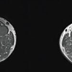
MRI activity changes in the sacroiliac joints (SIJs) that are consistent with SpA can also occurin individuals without SpA. Single lesions on MRI suggestive of inflammation have been reported in the SIJs in up to a quarter of young patients with mechanical back pain and healthy controls, studies have found.1

Normal variants can occur fairly frequently. Accessory sacroiliac joint has been seen in as many as 19% of subjects, iliosacral complex in 10%, and bipartite iliac bony plate in nearly 6%, Dr. Jurik said.

The Assessment of Spondyloarthritis International Society (ASAS) criteria for sacroiliitis is bone marrow edema seen using short inversion time recovery (STIR) or osteitis on T1 postcontrast images located in typical subchondral or periarticular bone areas—but the criteria don’t include structural changes, even though those can occur within the first two years of the disease.

“There is a need for revision of the ASAS MR criteria, with inclusion of structural changes,” Dr. Jurik said.

As for other techniques, whole-body MRI (WB-MRI) of the sacroiliac joints and spine can reliably detect inflammatory SIJs and spinal lesions in SpA patients, compared with conventional MRI, she said. There also might be value in imaging the anterior chest wall and enthesitis with this method, she said.

Studies have found that WB-MRI, including postcontrast sequence, detects significantly more areas of synovitis and enthesitis in SpA than through clinical means, except in the hands and feet, she said.2

Dr. Jurik said diffusion-weighted MRI, based on the movement of water protons, might be a way to diagnose early active sacroiliitis and includes the possibility of objectively quantifying inflammation using apparent diffusion coefficient values.

Page: 1 2 3 | Single Page
Share: 

Filed under:Axial SpondyloarthritisConditionsMeeting Reports Tagged with:EULARultrasonography

Related Articles

    Imaging in Ankylosing Spondylitis

    April 1, 2015

    MRI inflammation, fat and new bone formation in the sacroiliac joints, spine in patients with AS

    The Classification Challenge of Pediatric Spondylarthritis

    April 1, 2010

    Condition often confused with other disorders

    Musculoskeletal Ultrasound: A Valuable Tool for Diagnosing Rheumatic Illnesses

    Musculoskeletal Ultrasound: A Valuable Tool for Diagnosing Rheumatic Illnesses

    October 15, 2015

    Musculoskeletal (MSK) ultrasound is a valuable imaging modality for the practicing rheumatologist and provides an efficient tool with high diagnostic value in the evaluation of patients with musculoskeletal complaints. The use of MSK ultrasound has evolved in the U.S. due to the emergence of less-expensive, portable ultrasound units, which provide high-quality gray-scale and power Doppler…

    A Primer on Imaging in Myositis

    November 9, 2022

    In medicine, as in advertising, pictures can be worth a thousand words. From arthritis to vasculitis, imaging studies have been variably employed to aid in the diagnosis, treatment, risk stratification and prognostication of patients with rheumatic and musculoskeletal disorders. The same holds true with the idiopathic inflammatory myopathies (IIM), in which the clinical utility is…

  • About Us
  • Meet the Editors
  • Issue Archives
  • Contribute
  • Advertise
  • Contact Us
  • Copyright © 2025 by John Wiley & Sons, Inc. All rights reserved, including rights for text and data mining and training of artificial technologies or similar technologies. ISSN 1931-3268 (print). ISSN 1931-3209 (online).
  • DEI Statement
  • Privacy Policy
  • Terms of Use
  • Cookie Preferences