Video: Every Case Tells a Story| Webinar: ACR/CHEST ILD Guidelines in Practice

An official publication of the ACR and the ARP serving rheumatologists and rheumatology professionals

  • Conditions
    • Axial Spondyloarthritis
    • Gout and Crystalline Arthritis
    • Myositis
    • Osteoarthritis and Bone Disorders
    • Pain Syndromes
    • Pediatric Conditions
    • Psoriatic Arthritis
    • Rheumatoid Arthritis
    • Sjögren’s Disease
    • Systemic Lupus Erythematosus
    • Systemic Sclerosis
    • Vasculitis
    • Other Rheumatic Conditions
  • FocusRheum
    • ANCA-Associated Vasculitis
    • Axial Spondyloarthritis
    • Gout
    • Psoriatic Arthritis
    • Rheumatoid Arthritis
    • Systemic Lupus Erythematosus
  • Guidance
    • Clinical Criteria/Guidelines
    • Ethics
    • Legal Updates
    • Legislation & Advocacy
    • Meeting Reports
      • ACR Convergence
      • Other ACR meetings
      • EULAR/Other
    • Research Rheum
  • Drug Updates
    • Analgesics
    • Biologics/DMARDs
  • Practice Support
    • Billing/Coding
    • EMRs
    • Facility
    • Insurance
    • QA/QI
    • Technology
    • Workforce
  • Opinion
    • Patient Perspective
    • Profiles
    • Rheuminations
      • Video
    • Speak Out Rheum
  • Career
    • ACR ExamRheum
    • Awards
    • Career Development
  • ACR
    • ACR Home
    • ACR Convergence
    • ACR Guidelines
    • Journals
      • ACR Open Rheumatology
      • Arthritis & Rheumatology
      • Arthritis Care & Research
    • From the College
    • Events/CME
    • President’s Perspective
  • Search

Fellows’ Forum Case Report: Hemophagocytic Lymphohistiocytosis

Anita Laloo, MB, BS, MPH, German Pihan, MD, & Robert H. Shmerling, MD  |  Issue: August 2017  |  August 13, 2017

The patient was a 48-year-old woman who saw her primary care physician for a flu-like illness three months prior to admission. Her symptoms initially improved, but recurred one month later; she was treated symptomatically, and again symptoms resolved.

Two months later, she presented to an outside facility’s emergency department with fever to 103ºF, with associated chills, night sweats, headache, fatigue, abdominal pain and arthralgias, which became progressively more severe over one week. She reported having daily fever with headache, fatigue, malaise, myalgias and abdominal pain. There was no recent travel. Her episodes of abdominal discomfort were intermittent and primarily located in the epigastrium and left upper quadrant, without associated nausea, vomiting or diarrhea. Her arthralgias were localized to the bilateral metacarpophalangeal and proximal interphalangeal joints. Review of systems was otherwise negative.

ad goes here:advert-1
ADVERTISEMENT
SCROLL TO CONTINUE

Her past medical history was unremarkable, and she was up to date with routine health maintenance screening, including mammograms and pap smears. The only medication she was taking was acetaminophen for fever and arthralgias. She reported an allergy to penicillin. She had no family history of rheumatic disease. She lived with her husband and had four children. She had quit smoking 15 years earlier, but previously she’d had a 7.5 pack–year history of smoking.

At the outside hospital, she was found to be pancytopenic. This led to an extensive workup that was unrevealing, including tests for EBV, CMV, CMP, leptospira, parvovirus, toxoplasmosis, Babesia, anaplasma, Ehrlichia, Bartonella, influenza and Lyme disease, CT chest/abdomen/pelvis and a bone marrow biopsy. She was transferred to our medical center for additional evaluation.

ad goes here:advert-2
ADVERTISEMENT
SCROLL TO CONTINUE

Upon arrival, she was febrile to 102.2ºF, with a heart rate of 100 beats per minute, blood pressure of 90–100/40–50, and a respiratory rate of 24 with normal oxygen saturation on room air. She was pale in appearance. Her abdomen was soft with tenderness in the left upper quadrant and epigastrium, and the spleen was palpable. Her musculoskeletal exam revealed tenderness at the metacarpophalangeal and proximal interphalangeal joints of both hands, without swelling.

Laboratory studies included a white blood cell count of 2.4 K/µL, hemo­globin of 6.7 g/dL and platelet count of 84 K/µL. Her CRP was elevated to 90 mg/L and her ESR was 33 mm/h. Her ferritin was 543 ng/mL and triglyceride was

325 mg/dL. The remainder of her labs were unremarkable. She had a negative serological workup for ANA, ANCA, RF, anti-CCP and antibodies to RNP, Jo-1, Ro and La. SPEP revealed no abnormalities.
Page: 1 2 3 4 5 6 | Single Page
Share: 

Filed under:ConditionsResearch Rheum Tagged with:case reportClinicalDiagnosisFellowsFellows Forumfeverhemophagocytic lymphohistiocytosisimmunodeficiencylymphomamalignancyoutcomepatient careResearchrheumatologysymptoms

Related Articles

    Macrophage Activation Syndrome

    December 1, 2010

    A review of diagnosis, treatment, and prognosis

    Diagnosing & Treating Hemophagocytic Lymphohistiocytosis in Adults

    June 15, 2020

    Primary hemophagocytic lymphohistiocytosis (HLH) is caused by genetic mutations and inherited syndromes; it therefore occurs in the pediatric age group. Secondary HLH, however, is more common in adults and is often triggered by other disease states, such as malignancies, chronic immuno­suppression, infections and autoimmune disease.1,2 Macrophage activation syndrome (MAS) is a subset of secondary HLH…

    Case Report: Adult-Onset Still’s Disease with Complications

    February 14, 2023

    The following report outlines a case of newly diagnosed adult-onset Still’s disease (AOSD) complicated by macrophage activation syndrome (MAS) in a previously healthy and active 32-year-old man who had emigrated from Africa to the U.S. Case A man with no prior medical history presented with acute-onset polyarthritis, fevers and fatigue that began one month previously….

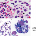
    Figure 1A–C: Arrows denote hemophagocytic histiocytes.

    Case Report: Too Many Activated Immune Cells in a 9-Month-Old Boy

    August 12, 2020

    Hemophagocytic lymphohistiocytosis (HLH) is a life-threatening disease of immune dysregulation characterized by unchecked inflammatory responses leading to end-organ dysfunction. Primary HLH results from inherited mutations that impair the capacity for immune regulation; secondary HLH arises from the inappropriate response to an immune stimulus, such as infection, malignancy or autoimmunity. What is less well known is…

  • About Us
  • Meet the Editors
  • Issue Archives
  • Contribute
  • Advertise
  • Contact Us
  • Copyright © 2025 by John Wiley & Sons, Inc. All rights reserved, including rights for text and data mining and training of artificial technologies or similar technologies. ISSN 1931-3268 (print). ISSN 1931-3209 (online).
  • DEI Statement
  • Privacy Policy
  • Terms of Use
  • Cookie Preferences