Video: Knock on Wood| Webinar: ACR/CHEST ILD Guidelines in Practice
fa-facebookfa-linkedinfa-youtube-playfa-rss

An official publication of the ACR and the ARP serving rheumatologists and rheumatology professionals

  • Conditions
    • Axial Spondyloarthritis
    • Gout and Crystalline Arthritis
    • Myositis
    • Osteoarthritis and Bone Disorders
    • Pain Syndromes
    • Pediatric Conditions
    • Psoriatic Arthritis
    • Rheumatoid Arthritis
    • Sjögren’s Disease
    • Systemic Lupus Erythematosus
    • Systemic Sclerosis
    • Vasculitis
    • Other Rheumatic Conditions
  • FocusRheum
    • ANCA-Associated Vasculitis
    • Axial Spondyloarthritis
    • Gout
    • Lupus Nephritis
    • Psoriatic Arthritis
    • Rheumatoid Arthritis
    • Systemic Lupus Erythematosus
  • Guidance
    • Clinical Criteria/Guidelines
    • Ethics
    • Legal Updates
    • Legislation & Advocacy
    • Meeting Reports
      • ACR Convergence
      • Other ACR meetings
      • EULAR/Other
    • Research Rheum
  • Drug Updates
    • Analgesics
    • Biologics/DMARDs
  • Practice Support
    • Billing/Coding
    • EMRs
    • Facility
    • Insurance
    • QA/QI
    • Technology
    • Workforce
  • Opinion
    • Patient Perspective
    • Profiles
    • Rheuminations
      • Video
    • Speak Out Rheum
  • Career
    • ACR ExamRheum
    • Awards
    • Career Development
  • ACR
    • ACR Home
    • ACR Convergence
    • ACR Guidelines
    • Journals
      • ACR Open Rheumatology
      • Arthritis & Rheumatology
      • Arthritis Care & Research
    • From the College
    • Events/CME
    • President’s Perspective
  • Search

Muscle Pathology for the Rheumatologist

Samantha C. Shapiro, MD  |  Issue: July 2022  |  June 17, 2022

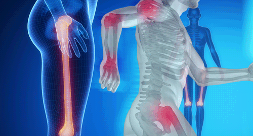
ORLANDO—In rheumatology, consults for muscle weakness and elevated creatinine kinase are common. The differential in these cases is broad, and when clinical history, laboratory studies and noninvasive testing fail to reveal a diagnosis, muscle biopsy can add valuable information. But to best use this information, we need to understand a bit about muscle pathology.

At the 2022 ACR Education Exchange, April 28–May 1, Peter Pytel, MD, professor of pathology, Department of Pathology, University of Chicago, shared his expertise.

ad goes here:advert-1
ADVERTISEMENT
SCROLL TO CONTINUE

Normal Muscle

Dr. Pytel began his talk with a review of normal muscle morphology. Normal skeletal muscle is composed of muscle fibers (cells) with peripheral nuclei and uniform size and cytoplasmic staining. Single muscle fibers are enwrapped in endomysium. Many muscle fibers are grouped into fascicles, which are enwrapped in perimysium. Normal fascicles don’t have visible connective tissue or inflammatory cells.

Muscle Disease

Dr. Pytel

When assessing muscle tissue, pathologists first look for myopathic changes: muscle fiber necrosis and regeneration. “First, we look for muscle fiber necrosis and atrophy that points to a myopathic process. Then, we use an algorithmic approach to better understand what’s going on,” Dr. Pytel said. “We look for inflammation, vacuoles and inclusions. We look for fat replacement and fibrosis that occur when muscle’s capacity to regenerate is overwhelmed, indicating chronicity. Finally, we use various staining techniques to refine our understanding further.”1

ad goes here:advert-2
ADVERTISEMENT
SCROLL TO CONTINUE

Generally, inflammatory myopathy—the term used to refer to autoimmune processes affecting skeletal muscle—is inflammation plus myopathy. However, some processes lack visible inflammatory infiltrates, such as immune-mediated necrotizing myopathy.

“Other disease processes, such as infection [e.g., trichinosis] and dystrophies [e.g., dysferlin deficiency], can exhibit inflammation too, so it’s important to consider the clinical context,” Dr. Pytel said.

Autoimmune Inflammatory Myopathies

Dr. Pytel shared pearls of wisdom specific to the autoimmune inflammatory myopathies we treat most frequently.

Regarding dermatomyositis, the hallmark feature is perifascicular atrophy. When viewed under the microscope, muscle fibers near the edges of fascicles appear shrunken compared with healthy fibers at the center. Perimysial inflammation (e.g., around the fascicles) is also present.  

“The perifascicular atrophy is far worse than the muscle necrosis,” Dr. Pytel said. “As for antisynthetase syndrome, add prominent necrotizing changes to the perifascicular atrophy we see in [dermatomyositis].”

When it comes to inclusion body myositis, the protracted clinical course is reflected in the biopsy. “We often see features of chronicity, like fibrosis and fatty replacement, that disrupts the normal fascicular architecture,” Dr. Pytel said.

Inflammation involves the endomysium (i.e., around individual muscle fibers in the fascicles), or the muscle fibers themselves. The rimmed vacuoles for which inclusion body myositis is named are best observed with modified Gomori trichome stain. Of note, rimmed vacuoles may also be seen in other neuromuscular disorders.2

Immune-mediated necrotizing myopathy, which is most commonly associated with anti-signal recognition particle (anti-SRP) and anti-3-hydroxy-3-methylglutaryl coenzyme A reductase (anti-HMGCR) antibodies, is characterized by random atrophy (e.g., mixed necrotic and regenerating fibers) and a relative paucity of inflammation.

“Compare this with [dermatomyositis], where the pattern is clearly perifascicular,” Dr. Pytel said.

Inflammatory myopathies have distinct but overlapping patterns, so when morphologic features overlap, pathologists can’t always offer a definitive diagnosis.

Don’t Forget the Drugs

It’s important to remember that medications can also cause muscle injury, and many of these toxicities have specific histologic findings. Example: Chronic steroids cause type 2 muscle fiber atrophy. Hydroxychloroquine causes a vacuolar myopathy with lysosomal aggregates visible with staining and curvilinear bodies visible on electron microscopy.

What’s the Diagnosis?

Inflammatory myopathies have distinct but overlapping patterns, so when morphologic features overlap, pathologists can’t always offer a definitive diagnosis.3

“In cases that aren’t clear-cut, immunohistochemical stains can provide additional information, and we’re discovering more of these each year,” Dr. Pytel said. “That’s why polymyositis is becoming a vanishingly small entity.”4

What about biopsies with mild myopathic changes? “A lot depends on clinical correlation,” Dr. Pytel said. “Is the biopsy representative of what led you to do the biopsy in the first place? A muscle biopsy with mild myopathic changes but no inflammatory infiltrates could be due to patchy distribution of disease and biopsy sampling error. Or perhaps it’s [immune-mediated necrotizing myopathy], in which case the absence of inflammation is expected. I’d also consider metabolic myopathy, dystrophies or toxic-metabolic damage. The clinical history is what provides the answers, so the more info you include with the specimen, the better.”

Summary

Dr. Pytel shared high-yield information that can help us better understand muscle pathology reports. Muscle biopsy may not always yield a definitive diagnosis due to morphologic overlap among myopathies. But when provided with ample clinical context, pathologists can refine their interpretations and provide information that allows us to better treat our patients.


Samantha C. Shapiro, MD, is an academic rheumatologist and an affiliate faculty member of the Dell Medical School at the University of Texas at Austin. She received her training in internal medicine and rheumatology at Johns Hopkins University, Baltimore. She is also a member of the ACR Insurance Subcommittee.

References

  1. Cai C, Anthony DC, Pytel P. A pattern-based approach to the interpretation of skeletal muscle biopsies. Mod Pathol. 2019 Apr;32(4):462–483.
  2. Jongen PJH, ter Laak HJ, Stadhouders AM. Rimmed basophilic vacuoles and filamentous inclusions in neuromuscular disorders. Neuromuscul Disord. 1995 Jan;5(1):31–38.
  3. Allenbach Y, Benveniste O, Goebel HH, et al. Integrated classification of inflammatory myopathies. Neuropathol Appl Neurobiol. 2017 Feb;43(1):62–81.
  4. Uruha A, Goebel HH, Stenzel W. Updates on the immunopathology in idiopathic inflammatory myopathies. Curr Rheumatol Rep. 2021 Jul 1;23(7):56.

Page: 1 2 3 | Multi-Page
Share: 

Filed under:ConditionsMeeting ReportsMyositisOther ACR meetings Tagged with:ACR Education Exchangedermatomyositis (DM)inflammatory myopathymuscleMusculoskeletal

Related Articles
    AJPhoto / Science Source

    Tips for Diagnosing Metabolic Myopathies

    September 17, 2019

    AJPhoto / Science Source When evaluating patients with possible myopathic symptoms, rheumatologists must consider a rare, but important, group of inherited disorders: the metabolic myopathies. However, their diagnosis often remains a challenge. Early recognition of these primary metabolic myopathies is essential to help prevent disease morbidity and mortality from rhabdomyolysis. Here, we focus on the…

    Diagnosis: Myopathy

    July 1, 2009

    Presentation and evaluation of metabolic causes

    New Tools for Myositis Diagnosis, Classification & Management

    April 15, 2019

    courtesy ACR Daily News CHICAGO—At Hot Topics in Myositis, a session at the 2018 ACR/ARHP Annual Meeting, three experts discussed new classification criteria for idiopathic inflammatory myopathies (IIM) and offered practical primers on overlap myositis conditions and inclusion body myositis (IBM). New Myositis Classification Criteria After a 10-year development process, the new EULAR/ACR Classification Criteria…

    Fellows’ Forum Case Report: Necrotizing Autoimmune Myopathy

    December 18, 2017

    Necrotizing autoimmune myopathy (NAM) is a relatively recently discovered subgroup of inflammatory myopathies. NAM is characterized by predominant muscle fiber necrosis and regeneration with little or no inflammation.1 One subgroup of NAM is 3-hydroxy-3-methylglutaryl-CoA reductase antibody (HMGCR Ab)-related immune-mediated necrotizing myopathy (IMNM), which occurs (rarely) after statin exposure, with a rough incidence of two per…

  • About Us
  • Meet the Editors
  • Issue Archives
  • Contribute
  • Advertise
  • Contact Us
fa-facebookfa-linkedinfa-youtube-playfa-rss
  • Copyright © 2025 by John Wiley & Sons, Inc. All rights reserved, including rights for text and data mining and training of artificial technologies or similar technologies. ISSN 1931-3268 (print). ISSN 1931-3209 (online).
  • DEI Statement
  • Privacy Policy
  • Terms of Use
  • Cookie Preferences