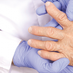
Imaging is not available in all settings, and Dr. van der Helm-van Mil explains they wanted a set of risk criteria that could be widely employed. Moreover, they designed the risk stratification tools to put comparatively more statistical weight on clinical and serological factors compared to imaging. But they also added the second and fuller risk criteria including MRI features that could be used for increased accuracy, if desired.
In the pooled analysis, 17% of patients had progressed to inflammatory arthritis by one year. Patients could be distributed into low risk of progression (<25% risk), intermediate risk (25–75%) or high risk (75% or higher). Including the use of MRI along with clinical and serological variables, 85% of patients fell into the lowest risk group, and 4% fell into the high-risk group.
For future study design, a cutoff point of 10 or greater points in the simplified version of the risk stratification criteria would yield a sensitivity and specificity of greater than 75% (or 12 or greater in the version including MRI). However, the committee did not recommend a specific reference risk stratification score for use in future trials. Dr. Deane pointed out that depending on the specific pre-RA study (e.g., agents with lower vs. higher side effect profile), researchers may use different cutoff points for inclusion of individuals.
Imaging Caveats
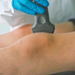
The lack of inclusion of ultrasound in the final risk stratification criteria was an important source of discussion among the committee members. Ultrasound’s lower cost and greater availability provide advantages over MRI. Moreover, although ultrasound did not emerge as providing additional predictive power in this specific pooled analysis, several papers in different international cohorts have shown that it may be predictive of progression to RA, including work by Dr Mankia and colleagues.11-13
Dr. Mankia notes that bone erosions seen via ultrasound have been highly predictive of progression in previous cohort studies.11-13 MRI is more sensitive for inflammation than ultrasound, particularly tenosynovitis, which may also have been part of why it improved predictive capacity in this analysis, even though ultrasound did not.
Dr. Mankia argues that ultrasound may still have utility in this population, and future risk stratification might ultimately include it. “In clinical practice, ultrasound helps us understand why people present with pain and stiffness,” he says, “because we can see inflammatory changes on the scan. If we do see those changes, we will likely monitor that patient more closely, because it is a sign that they may be on the way towards developing RA imminently.”