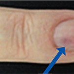
Video: Every Case Tells a Story| Webinar: ACR/CHEST ILD Guidelines in Practice

An official publication of the ACR and the ARP serving rheumatologists and rheumatology professionals

  • Conditions
    • Axial Spondyloarthritis
    • Gout and Crystalline Arthritis
    • Myositis
    • Osteoarthritis and Bone Disorders
    • Pain Syndromes
    • Pediatric Conditions
    • Psoriatic Arthritis
    • Rheumatoid Arthritis
    • Sjögren’s Disease
    • Systemic Lupus Erythematosus
    • Systemic Sclerosis
    • Vasculitis
    • Other Rheumatic Conditions
  • FocusRheum
    • ANCA-Associated Vasculitis
    • Axial Spondyloarthritis
    • Gout
    • Psoriatic Arthritis
    • Rheumatoid Arthritis
    • Systemic Lupus Erythematosus
  • Guidance
    • Clinical Criteria/Guidelines
    • Ethics
    • Legal Updates
    • Legislation & Advocacy
    • Meeting Reports
      • ACR Convergence
      • Other ACR meetings
      • EULAR/Other
    • Research Rheum
  • Drug Updates
    • Analgesics
    • Biologics/DMARDs
  • Practice Support
    • Billing/Coding
    • EMRs
    • Facility
    • Insurance
    • QA/QI
    • Technology
    • Workforce
  • Opinion
    • Patient Perspective
    • Profiles
    • Rheuminations
      • Video
    • Speak Out Rheum
  • Career
    • ACR ExamRheum
    • Awards
    • Career Development
  • ACR
    • ACR Home
    • ACR Convergence
    • ACR Guidelines
    • Journals
      • ACR Open Rheumatology
      • Arthritis & Rheumatology
      • Arthritis Care & Research
    • From the College
    • Events/CME
    • President’s Perspective
  • Search

The Role Ultrasound Imaging Plays in Diagnosing Hemangiomas

Clara Lin, MD, RhMSUS  |  Issue: April 2022  |  April 15, 2022

Sonographic characteristics of heman­giomas can be variable. Keng et al. described sonographic findings of 43 soft tissue hemangiomas.3 Gray-scale findings of hemangiomas demonstrated that 46.5% of lesions had a well-defined margin. Heman­giomas do not have a true capsule, so the appearance of a rim or margin may be due to the difference in echotexture between the hemangioma and the surrounding tissue, as well as an acoustic interface between the lesion and surrounding tissues. Most lesions were heterogenous (86%) and hypoechoic (60.5%). In larger lesions, fluid levels and calcified phleboliths can be seen.

Doppler activity was present in 86% (37 of 43) of hemangiomas.3 A color Doppler enhancing maneuver (CDEM) can be helpful in lesions without Doppler activity. CDEM is performed by placing light compression on the mass or adjacent tissues to enhance blood flow. CDEM was performed in the six lesions without Doppler activity and four demonstrated Doppler activity after CDEM.

ad goes here:advert-1
ADVERTISEMENT
SCROLL TO CONTINUE

In Sum

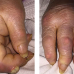
Hemangiomas are one of the most common tumors of childhood. Point-of-care ultrasound is a valuable tool to better evaluate deep lesions that are difficult to diagnose by physical exam. Most lesions are heterogeneous, hypoechoic and exhibit Doppler activity, so they can be recognized and distinguished by ultrasound from other causes of soft tissue nodules encountered in the rheumatology clinic.


Clara Lin, MD, RhMSUS, is a staff pediatric rheumatologist at Children’s Hospital Colorado, Aurora. Her clinical and research interests include ultrasound in rheumatology. She is also a board member of USSONAR, a society of rheumatologists, physicians and health professionals in North America who promote the use of musculoskeletal ultrasonography to advance the care of patients with rheumatic diseases.

ad goes here:advert-2
ADVERTISEMENT
SCROLL TO CONTINUE

References

  1. Darrow DH, Greene AK, Mancini AJ, Nopper AJ. Section on dermatology, section on otolaryngology-head & neck surgery and section on plastic surgery. Diagnosis and management of infantile hemangioma: Executive summary. Pediatrics. 2015 Oct;136(4):786–791.
  2. Drapé JL, Feydy A, Guerini H, et al. Vascular lesions of the hand. Eur J Radiol. 2005 Dec;56(3):331–343.
  3. Keng CY, Lan HHC, Chen CCC, et al. Soft tissue hemangiomas: High-resolution grayscale and color Doppler ultrasonographic features in 43 patients. J Med Ultrasound. 2008 Dec:16(3):223–230.

Page: 1 2 | Single Page
Share: 

Filed under:Conditions Tagged with:diagnostic imaginghemangiomasUltrasound

Related Articles
    Musculoskeletal Ultrasound: A Valuable Tool for Diagnosing Rheumatic Illnesses

    Musculoskeletal Ultrasound: A Valuable Tool for Diagnosing Rheumatic Illnesses

    October 15, 2015

    Musculoskeletal (MSK) ultrasound is a valuable imaging modality for the practicing rheumatologist and provides an efficient tool with high diagnostic value in the evaluation of patients with musculoskeletal complaints. The use of MSK ultrasound has evolved in the U.S. due to the emergence of less-expensive, portable ultrasound units, which provide high-quality gray-scale and power Doppler…

    Fellows’ Forum Case Report: Tophaceous Bullae

    August 13, 2017

    As a manifestation of hyperuricemia, inflammatory bullous lesions have rarely been described in the past century. A more classic presentation of hyperuricemia is acute inflammatory gouty arthritis, characterized by the deposition of monosodium urate crystals. Other complications of chronic untreated hyperuricemia may include polyarticular arthritis, tophus formation and possible chronic destructive lesions of the bone,…

    Sonographic Diagnosis of Knuckle Pads

    October 10, 2022

    The Case A 56-year-old white woman was evaluated for a one-year history of painless bumps on the dorsal aspect of the proximal interphalangeal (PIP) joints of both hands and suspected flexor tenosynovitis in her palms. On examination, small cystic nodules without erythema or tenderness were present on the dorsal aspect of several PIP joints (see…

    Figure 2: The nail bed shows some discoloration.

    3 Cases of Glomus Tumor—An Unusual Cause of Hand Pain

    May 13, 2016

    Hand and digit pain are common presenting symptoms to primary care physicians, rheumatologists, physiatrists and neurologists. There are many causes, but quality and location of the pain can be important clues to the diagnosis. Glomus tumors, neurovascular hamartomas of the glomus body, are an uncommon cause of hand pain. The glomus unit itself is a…

  • About Us
  • Meet the Editors
  • Issue Archives
  • Contribute
  • Advertise
  • Contact Us
  • Copyright © 2025 by John Wiley & Sons, Inc. All rights reserved, including rights for text and data mining and training of artificial technologies or similar technologies. ISSN 1931-3268 (print). ISSN 1931-3209 (online).
  • DEI Statement
  • Privacy Policy
  • Terms of Use
  • Cookie Preferences