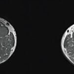
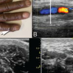
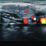
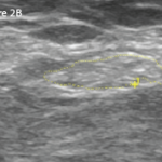
Video: Every Case Tells a Story| Webinar: ACR/CHEST ILD Guidelines in Practice

An official publication of the ACR and the ARP serving rheumatologists and rheumatology professionals

  • Conditions
    • Axial Spondyloarthritis
    • Gout and Crystalline Arthritis
    • Myositis
    • Osteoarthritis and Bone Disorders
    • Pain Syndromes
    • Pediatric Conditions
    • Psoriatic Arthritis
    • Rheumatoid Arthritis
    • Sjögren’s Disease
    • Systemic Lupus Erythematosus
    • Systemic Sclerosis
    • Vasculitis
    • Other Rheumatic Conditions
  • FocusRheum
    • ANCA-Associated Vasculitis
    • Axial Spondyloarthritis
    • Gout
    • Psoriatic Arthritis
    • Rheumatoid Arthritis
    • Systemic Lupus Erythematosus
  • Guidance
    • Clinical Criteria/Guidelines
    • Ethics
    • Legal Updates
    • Legislation & Advocacy
    • Meeting Reports
      • ACR Convergence
      • Other ACR meetings
      • EULAR/Other
    • Research Rheum
  • Drug Updates
    • Analgesics
    • Biologics/DMARDs
  • Practice Support
    • Billing/Coding
    • EMRs
    • Facility
    • Insurance
    • QA/QI
    • Technology
    • Workforce
  • Opinion
    • Patient Perspective
    • Profiles
    • Rheuminations
      • Video
    • Speak Out Rheum
  • Career
    • ACR ExamRheum
    • Awards
    • Career Development
  • ACR
    • ACR Home
    • ACR Convergence
    • ACR Guidelines
    • Journals
      • ACR Open Rheumatology
      • Arthritis & Rheumatology
      • Arthritis Care & Research
    • From the College
    • Events/CME
    • President’s Perspective
  • Search

Articles tagged with "Ultrasound"

Clockwise from top: Image A shows the periungual ulceration (arrow) of the fifth digit, as well as the prior amputation of the second digit. Image B shows a longitudinal ultrasound of the palmar aspect of the ulnar right wrist. In the center of the image, the ulnar artery is in view and color Doppler flow is visualized within the vessel until there is a reversal of flow demonstrated by color change, followed by severe attenuation of flow distally. Distal to the cessation of flow, the vessel appears hypoechoic due to proliferation of the intima to the point of occluding the lumen. Image C shows a transverse view at the level of white line in Image B, with endothelial proliferation seen around a central lumen of the artery (a), next to the vein (v) and ulnar nerve (n). Image D, shows a transverse view at the level of the black line in Image B, where the arterial lumen (a) is occluded.

Insights Into Ulnar Artery Occlusion in Systemic Sclerosis

Devin M. Driscoll, MD, Andreea M. Bujor, MD, PhD, & Eugene Y. Kissin, MD, Rhmsus  |  April 17, 2021

A 51-year-old man with a history of limited systemic sclerosis with Raynaud’s phenomenon and pulmonary hypertension being treated with tadalafil and macitentan presented to a clinic with ulceration of his right pinkie. The patient had injured the finger two months earlier. He reported poor healing and the presence of a persistent ulcer since the injury….

Ultrasound Provides Insights into Immune Checkpoint Inhibitor‐Induced Inflammatory Arthritis

Elizabeth Hofheinz, MPH, MEd  |  October 12, 2020

Ultrasound may provide unique insights into the effects of immune checkpoint inhibitors on the human body beyond the immune system. Research suggests synovitis and inflammatory tendon involvement are commonly seen in patients with immune checkpoint inhibitor-induced inflammatory arthritis.

A transverse view of the ulnar groove in full elbow extension. The red arrow indicates the advancing edge of the MHTr.

Recurrent Medial Elbow Pain Following Successful Tommy John Surgery

Mark H. Greenberg, MD, RMSK, RhMSUS, A. Lee Day, MD, RMSK, James W. Fant Jr., MD, & Christopher G. Mazoue, MD  |  August 12, 2020

A 27-year-old, left-handed man was referred to our ultrasound clinic for left elbow pain. History The patient had been a pitcher on a Minor League Baseball team. Two years before, he developed sudden, severe medial elbow pain while pitching in a game. The pain was associated with some tingling down the left medial forearm. The…

Ultrasound in Rheumatology—Past, Present & Future

Jason Liebowitz, MD, FACR  |  July 15, 2020

For most rheumatologists, the key elements of the physical exam—inspection, palpation, percussion and auscultation—have long been second nature, but a fifth modality has grown in importance with respect to making the correct diagnosis: ultrasound. From evaluating for Doppler signal and additional findings indicative of synovitis to identifying bony erosions, chondrocalcinosis, tophi and other articular and…

Right: The same view as 2A, with the common peroneal nerve outlined in yellow with a cross-sectional area of 21 mm2.

Case Report: Ultrasound Reveals Cause of Post-Arthroplasty Knee Pain

Mark H. Greenberg, MD, RMSK, RhMSUS, Elijah Mitcham, MD, Prem Patel, James W. Fant Jr., MD, & Frank R. Voss, MD  |  April 15, 2020

A 65-year-old woman was referred by an orthopedist to a rheumatologist for left knee pain. Previously, in 2014, she underwent left total knee arthroplasty (TKA) for severe osteoarthritis in a different institution. Following the procedure, she experienced severe chronic anterolateral knee pain at rest, exacerbated by walking. Because she was rendered wheelchair bound and required…

How to Diagnose Upper Extremity Injuries

Mike Fillon  |  February 13, 2020

ATLANTA—Two ways to investigate injuries to the upper extremities are by in-depth physical examinations and ultrasound. In a Clinical Practice session at the 2019 ACR/ARP Annual Meeting, Anatomy: Correlating Physical Exam and Ultrasound in Common Sports Injuries of the Upper Extremity, Carlin Senter, MD, FACP, associate professor of primary care sports medicine at the University…

According to MRI, Ultrasound Guidance Does Not Improve Treat-to-Target in RA

Reuters Staff  |  February 5, 2020

NEW YORK (Reuters Health)—Ultrasound guidance does not improve the effectiveness of treat-to-target therapy in rheumatoid arthritis (RA), new findings confirm. “Incorporating ultrasound information in treatment decisions did not lead to reduced MRI inflammation or less structural damage compared with a conventional treatment strategy,” Dr. Ulf Sundin of Diakonhjemmet Hospital, Oslo, and colleagues write in Rheumatology….

Coding Corner Answer: Is This Shoulder Ultrasound Billable?

From the College  |  September 17, 2019

Take the challenge. Answer: No, if billed as incident-to for the NP. Note: The patient’s current insurance carrier is Blue Cross Blue Shield of Massachusetts (BCBS of MA). Under BCBS of MA, nurse practitioners and nurse practitioner primary care providers are not allowed to perform or interpret radiology services. Therefore, the physician would have to…

Coding Corner Question: Is This Shoulder Ultrasound Billable?

From the College  |  September 17, 2019

A 32-year-old male patient with rheumatoid arthritis affecting multiple joints without rheumatoid factor returns to the office for a follow-up visit with the nurse practitioner (NP). The patient states his overall pain has improved since his previous visit, although he rates the severity of his right shoulder and right wrist pain at a 5 on…

Advanced Practice Providers Should Learn Musculoskeletal Ultrasound

Heather Benham, DNP, RN, CPNP, RhMSUS, on behalf of the ARP Practice Committee   |  June 17, 2019

I have worked as an advanced practice provider (APP) in pediatric rheumatology for nearly 16 years. My collaborating physicians have allowed me to function to the full extent of my scope of practice while allowing me to develop professionally within the subspecialty. My latest endeavor has been learning how to perform musculoskeletal ultrasound (MSUS) and…

  • « Previous Page
  • 1
  • 2
  • 3
  • 4
  • …
  • 8
  • Next Page »
  • About Us
  • Meet the Editors
  • Issue Archives
  • Contribute
  • Advertise
  • Contact Us
  • Copyright © 2025 by John Wiley & Sons, Inc. All rights reserved, including rights for text and data mining and training of artificial technologies or similar technologies. ISSN 1931-3268 (print). ISSN 1931-3209 (online).
  • DEI Statement
  • Privacy Policy
  • Terms of Use
  • Cookie Preferences