CHICAGO—How magnetic resonance imaging (MRI) is deployed for the diagnosis of axial spondyloarthritis (axSpA) can make a huge difference—much more important than clinical presentation and even other objective criteria, said Walter Maksymowych, MB ChB, professor of medicine at the University of Alberta, Edmonton, Canada, in a session at ACR Convergence 2025.
“Clinical features aren’t particularly helpful in making a diagnosis, and we, in fact, rely a lot more on the objective manifestations,” Dr. Maksymowych noted. “But even there, an elevated CRP (C-reactive protein) [is seen] in only 40% of patients. So if you’re relying on the CRP, that’s not going to get you very far.”
His review of the diagnosis of axSpA came in a session that also included updates on juvenile axSpA and mimics that clinicians have to keep in mind as they assess their patients.
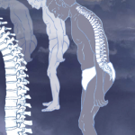
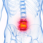
MRI Findings Crucial to Diagnosis

Dr. Maksymowych
Most integral to the diagnosis of axSpA are findings on magnetic resonance imaging (MRI), Dr. Maksymowych said. As illustrated by the recent CLASSIC study, the specifications of the MRI are particularly important.1
The CLASSIC study set out to validate the performance of the Assessment of SpondyloArthritis International Society (ASAS) classification criteria in a worldwide cohort, ultimately enrolling about 1,000 patients seen consecutively by a rheumatologist after at least three months of undiagnosed back pain. The researchers found that MRI was much better than most other measures at capturing axSpA, with 81.9% of axSpA diagnoses having an MRI indicative of the disease among local readings of the imaging and 61.6% of central readings.
The MRI protocol used in CLASSIC was T1 semi-coronal, STIR semi-coronal, STIR axial and a T1-weighted, fat-suppressed, 3D gradient echo, thin-slice sequence that is erosion-sensitive. These will help identify inflammatory as well as structural lesions and will allow bone marrow edema to be precisely located, Dr. Maksymowych said. It is the synthesis of all of these sequences that brings the picture into focus, he noted.
The upshot, he said, is that there is now a specific protocol that you “can take to a local radiologist and say, ‘I want you to do a certain acquisition protocol for diagnostic evaluation of the SI [sacroiliac] joint.’ This is the type of MRI that I want to see in my clinical practice.”
In the case of many patients with lower back pain who don’t respond to non-steroidal anti-inflammatory drugs, clinicians reach a pivotal moment when they need to decide whether to start someone on a biologic. “Now you’re struggling to decide, am I going to go into something that’s potentially lifelong therapy?” he said. “The imaging is going to be crucial to make that decision.”
Axial Juvenile Spondyloarthritis

Dr. Weiss
Pamela Weiss, MD, clinical research director of the Rheumatology Division at Children’s Hospital of Philadelphia, reviewed some of the subtleties and challenges in axial juvenile spondyloarthritis (axJSpA).
It’s helpful to think of axJSpA as having a rule of 20, she said. About 20% of SpA is juvenile onset, about 20% of juvenile idiopathic arthritis (JIA) is JSpA, about 20% of JSpA cases involve axial disease, and about 20% of JSpA cases fail to achieve remission within five years.
Dr. Weiss cautioned that, when reading pediatric MRI images of the sacroiliac joint, a bright signal often does not indicate disease; the maturing SI joint tends to appear this way. A handy way to tell the difference, she said, is that if it looks like the whiteness around the edge of the joint could have been neatly drawn in with a Sharpie pen, “it’s probably just maturational signal.”
AxJSpA classification criteria published last year include seven domains with two or three levels in each domain, scored on a scale of 100, with 55 the threshold for a diagnosis.2 MRI findings are given the most weight in the scoring, and an online calculator is available, Dr. Weiss said.3
Key differences between the juvenile and the adult criteria are that back, hip or groin pain can satisfy the requirements for juveniles, but only back pain is relevant in the adult criteria because young people can often have different ways of describing their pain, she said. Also, among juveniles, both imaging and clinical criteria must be met, rather than either one for adults.
Treatment options are much more limited for the JSpA population, Dr. Weiss noted. Research has also found that juvenile onset of axSpA tends to lead to fewer functional issues with the spine as people age, compared with adult-onset disease, but more hip functional issues compared with adult-onset disease.4
Mimics of axSpA

Dr. Gensler
Lianne Gensler, MD, director of the University of California, San Francisco Ankylosing Spondylitis Clinic, drew attention to the many mimics that can initially appear to be possible axSpA.
Pregnancy is an important contributor to mimics of sacroiliitis, Dr. Gensler said. Women can have bone marrow edema from stress fractures during pregnancy that can persist for a year after they’ve given birth, but it might appear to be sacroiliitis. Low-dose computed tomography scans can provide additional clarity that can be highly useful. “Just interpret those [imaging] results with caution because it may be sacroiliitis, but it could also just be the pregnancy-related changes,” she noted. Osteitis condensans ilii and degenerative disease are two axSpA mimics that can be brought on by pregnancy.
Malignancies with spine infiltration, such as B cell lymphoma or acute lymphocytic leukemia, can also masquerade as axSpA, Dr. Gensler said.
Other mimics to watch out for include diffuse idiopathic skeletal hyperostosis (DISH); Scheurmann’s disease, involving an abnormal and excessive curvature of the spine; and Bertolotti syndrome, involving an extra bone between the last lumbar bone and the sacrum, Dr. Gensler said. Clinicians should also be aware of the possibility of sacral insufficiency fractures, spinal fractures and infections.
Dr. Gensler said clinicians should keep a list of pointers in mind so as not to fall into the axSpA trap. First, inflammatory back pain does not necessarily equal axSpA, even when other suggestive features are present; clinical context and concordant imaging are needed. Second, clinicians should read MRIs “like a detective,” on the lookout for other diagnoses. Third, a stress reaction and cortical irregularity don’t necessarily add up to the presence of sacroiliitis. Fourth, clinicians should have a list of diagnoses they “can’t miss,” including infections, malignancies and fractures.
Lastly, she said, it’s important to keep in mind that Hickam’s dictum—that “patients can have as many diseases as they damn well please”—often supersedes Occam’s razor, the idea that the simplest explanation is usually the best.
“The law of simplicity to explain everything,” Dr. Gensler said, “doesn’t always work.”
Thomas R. Collins is a freelance medical writer based in Florida.
References
- Maksymowych WP, van der Heijde D, Caplan L, et al. The Assessments in Spondyloarthritis International Society (ASAS) and Spondyloarthritis Research and Treatment Network (SPARTAN) Revised Classification Criteria for Axial Spondyloarthritis: Development and validation in the classification of axial SpA inception cohort study [abstract 0854]. Arthritis Rheumatol. 2025;77(suppl 9).
- Weiss PF, Brandon TG, Aggarwal A, et al. Classification criteria for axial disease in youth with juvenile spondyloarthritis. Arthritis Rheumatol. 2024 Dec;76(12):1797–1808.
- Children’s Hospital of Philadelphia. Axial Juvenile [PF3] Spondyloarthritis (AxJSpA) Classification Criteria Calculator. https://redcap.chop.edu/surveys/?s=D3CXKYTRL4M87XM8. Accessed October 31, 2025.
- Gensler LS, Ward MM, Reveille JD, et al. Clinical, radiographic and functional differences between juvenile-onset and adult-onset ankylosing spondylitis: results from the PSOAS cohort. Ann Rheum Dis. 2008 Feb;67(2):233–237.


