 Jointly sponsored by the European League Against Rheumatism (EULAR) and the ACR, the 2019 classification criteria for systemic lupus erythematosus (SLE) were published in a special article in the September issue of Arthritis & Rheumatology.1 The criteria offer improved sensitivity and specificity compared with the 1997 update of the 1982 ACR criteria and the 2012 Systemic Lupus International Collaborating (SLICC) criteria.2,3 Additionally, they more accurately reflect the current tests used to clinically diagnose SLE and are a “simple, directed and highly accurate method for classifying SLE,” write the authors.
The 2019 EULAR/ACR criteria are designed to identify SLE patients for inclusion in clinical trials and research studies—not for clinical diagnosis. They were produced by the largest collaboration of international lupus experts and patients to date, says co-investigator Sindhu Johnson, MD, PhD, clinician-scientist, University Health Network and Sinai Health Systems, and associate professor of medicine at the University of Toronto, Canada.
“This project involved more than 200 investigators from a variety of specialties and methodologies, as well as more than 4,000 patients. Lupus patients were involved in the development of the criteria and the quantitative validation process,” says Dr. Johnson.
Early SLE Patients Included
Although the 2012 SLICC criteria addressed some of the shortcomings of the previous ACR criteria (e.g., the addition of mucocutaneous and neuropsychiatric manifestations, hypocomplementemia and anti-nuclear antibody [ANA] tests) and offered more refined criteria definitions, their specificity was lower than the 1997 ACR criteria. The 2019 EULAR/ACR criteria have a sensitivity of 96.1% and a specificity of 93.4% when tested in the validation cohort. Although the 1997 ACR classification criteria have the same specificity of 93.4%, they have a sensitivity of only 82.8%. The SLICC criteria have a slightly higher sensitivity than the new set, or 96.7%, but a specificity of only 83.7%.
Additionally, both the 1997 and 2012 criteria perform better in patients with longstanding SLE than those with early disease, and new-onset SLE patients should now have a greater representation in trials. The early SLE sample populations were purposely included in each cohort in the four phases of development of the 2019 criteria.
In the first phase, ANA was evaluated as an entry criterion—the first hurdle to classify a patient as having SLE—through systematic review and meta-regression of the literature. Then, criteria were generated through an international Delphi exercise, evaluation of a cohort of early SLE patients and a patient survey. In phase 2, the criteria were reduced in another Delphi exercise and nominal group technique exercises. Next, criteria were defined and weighted on the basis of their performance and the results of a multi-criteria decision analysis. The criteria weights and threshold scores were refined in a new cohort of 1,001 patients. Then the set was validated by comparing its performance with that of the previous criteria in another cohort of 1,270 patients.
Significant Updates
The 2019 criteria include significant changes from the previous criteria, says Dr. Johnson. First, any patient must have had at least one positive ANA test as an obligatory criterion. If the patient has not had a positive ANA test or has been consistently ANA negative, they cannot be classified as having SLE no matter how many other criteria they may fulfill. Although a patient who is ANA negative could be correctly diagnosed with lupus, this is uncommon, so classification criteria don’t need to account for this.
“ANAs were recognized as an important criterion for the classification of lupus in the previous criteria and in clinical practice. Rheumatologists use ANA as a screening test for lupus. Using ANA as an entry criterion [to SLE classification] is a better reflection of current clinical practice,” she says.
After a patient fulfills this criterion, users then determine how many of 22 other SLE criteria the patient fulfills (see Table 1). These criteria represent seven clinical domains (i.e., constitutional, hematologic, neuropsychiatric, mucocutaneous, serosal, musculoskeletal and renal) and three immunologic domains (i.e., antiphospholipid antibodies, complement proteins and SLE-specific antibodies). Each criterion is weighted from 2 to 10 for its specific applicability to lupus. (Note: Refer to the full article in Arthritis & Rheumatology for the weights and all supporting references to the literature.1)
The scoring tool includes these directives:
- Any criterion that occurred on at least one occasion is sufficient to qualify;
- SLE classification requires at least one clinical criterion and 10 or more points;
- Criteria do not have to occur simultaneously; and
- Within each domain, only the criterion with the highest weight counts toward the total score.
The ACR will be adding the criteria to its mobile guideline/criteria app, which will give researchers quick access to the criteria for classification purposes.
| Table 1: The Criteria Defined1 | |
| Anti-nuclear antibodies (ANA) |
ANA at a titer of ≥1:80 on HEp-2 cells or an equivalent positive test at least once. Testing by immunofluorescence on HEp-2 cells or a solid-phase ANA screening immunoassay with at least equivalent performance is highly recommended |
| Fever |
Temperature >38.3°C |
| Leukopenia |
White blood cell count <4,000/mm³ |
| Thrombocytopenia |
Platelet count <100,000/mm³ |
| Autoimmune hemolysis |
Evidence of hemolysis, such as reticulocytosis, low haptoglobin, elevated indirect bilirubin, elevated LDH, AND positive Coombs’ (direct antiglobulin) test |
| Delirium |
Characterized by 1) change in consciousness or level of arousal with reduced ability to focus, 2) symptom development over hours to <2 days, 3) symptom fluctuation throughout the day, 4) either 4a) acute/subacute change in cognition (e.g., memory deficit or disorientation), or 4b) change in behavior, mood, or affect (e.g., restlessness, reversal of sleep/wake cycle) |
| Psychosis |
Characterized by 1) delusions and/or hallucinations without insight and 2) absence of delirium |
| Seizure |
Primary generalized seizure or partial/focal seizure |
| Non-scarring alopecia |
Non-scarring alopecia observed by a clinician† |
| Oral ulcers |
Oral ulcers observed by a clinician† |
| Subacute cutaneous OR discoid lupus |
Subacute cutaneous lupus erythematosus observed by a clinician:† Annular or papulosquamous (psoriasiform) cutaneous eruption, usually photodistributed If skin biopsy is performed, typical changes must be present (interface vacuolar dermatitis consisting of a perivascular lymphohistiocytic infiltrate, often with dermal mucin noted). OR discoid lupus erythematosus observed by a clinician:† Erythematous-violaceous cutaneous lesions with secondary changes of atrophic scarring, dyspigmentation, often follicular hyperkeratosis/plugging (scalp), leading to scarring alopecia on the scalp If skin biopsy is performed, typical changes must be present (interface vacuolar dermatitis consisting of a perivascular and/or periappendageal lymphohistiocytic infiltrate. In the scalp, follicular keratin plugs may be seen. In longstanding lesions, mucin deposition may be noted) |
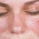
| Acute cutaneous lupus |
Malar rash or generalized maculopapular rash observed by a clinician† If skin biopsy is performed, typical changes must be present (interface vacuolar dermatitis consisting of a perivascular lymphohistiocytic infiltrate, often with dermal mucin noted. Perivascular neutrophilic infiltrate may be present early in the course) |
| Pleural or pericardial effusion |
Imaging evidence (such as ultrasound, x-ray, CT scan, MRI) of pleural or pericardial effusion, or both |
| Acute pericarditis |
≥2 of 1) pericardial chest pain (typically sharp, worse with inspiration, improved by leaning forward), 2) pericardial rub, 3) EKG with new widespread ST elevation or PR depression, 4) new or worsened pericardial effusion on imaging (such as ultrasound, x-ray, CT scan, MRI) |
| Joint involvement |
EITHER 1) synovitis involving 2 or more joints characterized by swelling or effusion OR 2) tenderness in 2 or more joints and at least 30 minutes of morning stiffness |
| Proteinuria >0.5 g/24 hours |
Proteinuria >0.5 g/24 hours by 24-hour urine or equivalent spot urine protein-to-creatinine ratio |
| Class II or V lupus nephritis on renal biopsy according to ISN/RPS 2003 classification>0.5 g/24 hours |
Class II: Class V: |
| Class III or IV lupus nephritis on renal biopsy according to ISN/RPS 2003 classification |
Class III: Class IV: |
| Positive antiphospholipid antibodies |
Anticardiolipin antibodies (IgA, IgG, or IgM) at medium or high titer (>40 APL, GPL, or MPL, or >the 99th percentile) or positive anti-β2GPI antibodies (IgA, IgG, or IgM) or positive lupus anticoagulant |
| Low C3 OR low C4 |
C3 OR C4 below the lower limit of normal |
| Low C3 AND low C4 |
Both C3 AND C4 below their lower limits of normal |
| Anti-dsDNA antibodies OR anti-Sm antibodies |
Anti-dsDNA antibodies in an immunoassay with demonstrated ≥90% specificity for SLE against relevant disease controls OR anti-Sm antibodies |
| * SLE = systemic lupus erythematosus; LDH = lactate dehydrogenase; CT = computed tomography; MRI = magnetic resonance imaging; EKG = electrocardiography; ISN = International Society of Nephrology; RPS = Renal Pathology Society; anti-β2GPI = anti–β2-glycoprotein I; anti-dsDNA = anti–double-stranded DNA. † This may include physical examination or review of a photograph. |
|
Weighted Scores
Weighting scores is a novel feature of the new criteria and improves their utility for lupus classification, says Dr. Johnson.
“Previously, all criteria were weighted equally, but in the modern era of criteria development and validation, weighting improves the results. Our weighting of SLE criteria is a better reflection of the way rheumatologists practice today,” she says. “For example, lupus nephritis is weighted more heavily than mouth ulcers. Any individual who is positive for ANA and has class III or IV lupus nephritis can be classified as having SLE. In our development process, international lupus experts all told us that class III or IV lupus nephritis is important for classification.”
Specificity to lupus adds weight to particular criteria, she says. Class III or IV renal involvement scores 10 points. Acute pericarditis, acute cutaneous lupus erythematosus (ACLE), joint involvement, anti-Sm and anti-dsDNA antibodies all weigh six points each. However, oral ulcers, fever, delirium, alopecia and antiphospholipid antibodies—all of which may be present in other conditions—weigh just two points each.
SLE is a heterogeneous and complex disease. Thus, clinical manifestations that are not likely caused by lupus should not be counted, notes Dr. Johnson.
“Fever is a novel criterion in these criteria. But it’s important for rheumatologists to rule out other causes for a patient’s fever and to count the fever only if it is attributable to lupus. Why do we count only one criterion in a domain? Because, in each domain, many criteria can be related to each other. It is important to count only the highest weight within a domain,” she says.
Some Novel Biomarkers Excluded
Many novel molecular biomarkers for SLE, such as increased circulating B lymphocyte stimulator, interferon-g-induced protein kD, monocyte chemoattractant protein 1, tumor necrosis factor-a, type 1 interferon signature and increased Th17 and plasma cell populations, were nominated for inclusion in the development process. However, these biomarkers were voted out of inclusion in the Delphi exercise due to insufficient evidence or limited global availability of tests for these biomarkers.
“There are groups in the U.S. and around the world who are doing very important work on biomarkers in lupus. But classification criteria must be feasible, validated and reliable at different sites worldwide. Some biomarkers are not accessible at many sites. In the future, I think we will move toward more molecular or biological classification of lupus, but we are not there yet,” Dr. Johnson says.
The classification criteria do not include a complete list of all the possible manifestations a rheumatologist may see in lupus. And the new criteria have a simplified list compared with the SLICC criteria, Dr. Johnson stresses.
“Diagnosis of lupus remains in the hands of an appropriately trained healthcare professional. It’s equally important to state that payers, especially in the U.S., should not deny appropriate therapies or reimbursement for those therapies in a patient who does not fulfill the classification criteria,” she says.
Susan Bernstein is a freelance journalist based in Atlanta.
References
- Aringer M, Costenbader K, Daikh D, et al. 2019 European League Against Rheumatism/American College of Rheumatology Classification Criteria for Systemic Lupus Erythematosus. Arthritis Rheumatol. 2019 Sep;71(9):1400–1412.
- Hochberg MC. Updating the American College of Rheumatology revised criteria for the classification of systemic lupus erythematosus. Arthritis Rheum. 1997 Sep;40(9):1725.
- Petri M, Orbai AM, Alarcon GS, et al. Derivation and validation of the Systemic Lupus International Collaborating Clinics classification criteria for systemic lupus erythematosus. Arthritis Rheum. 2012 Aug;64(8):2677–2686.