Video: Every Case Tells a Story| Webinar: ACR/CHEST ILD Guidelines in Practice

An official publication of the ACR and the ARP serving rheumatologists and rheumatology professionals

  • Conditions
    • Axial Spondyloarthritis
    • Gout and Crystalline Arthritis
    • Myositis
    • Osteoarthritis and Bone Disorders
    • Pain Syndromes
    • Pediatric Conditions
    • Psoriatic Arthritis
    • Rheumatoid Arthritis
    • Sjögren’s Disease
    • Systemic Lupus Erythematosus
    • Systemic Sclerosis
    • Vasculitis
    • Other Rheumatic Conditions
  • FocusRheum
    • ANCA-Associated Vasculitis
    • Axial Spondyloarthritis
    • Gout
    • Psoriatic Arthritis
    • Rheumatoid Arthritis
    • Systemic Lupus Erythematosus
  • Guidance
    • Clinical Criteria/Guidelines
    • Ethics
    • Legal Updates
    • Legislation & Advocacy
    • Meeting Reports
      • ACR Convergence
      • Other ACR meetings
      • EULAR/Other
    • Research Rheum
  • Drug Updates
    • Analgesics
    • Biologics/DMARDs
  • Practice Support
    • Billing/Coding
    • EMRs
    • Facility
    • Insurance
    • QA/QI
    • Technology
    • Workforce
  • Opinion
    • Patient Perspective
    • Profiles
    • Rheuminations
      • Video
    • Speak Out Rheum
  • Career
    • ACR ExamRheum
    • Awards
    • Career Development
  • ACR
    • ACR Home
    • ACR Convergence
    • ACR Guidelines
    • Journals
      • ACR Open Rheumatology
      • Arthritis & Rheumatology
      • Arthritis Care & Research
    • From the College
    • Events/CME
    • President’s Perspective
  • Search

A Case of Nodular Rash & Painful Joints

Vania Lin, MD, MPH, Rebecca Johnson, MD, & Lisa Suter, MD  |  Issue: October 2022  |  October 10, 2022

He denied any of the following symptoms: sore throat, cough, nasal congestion, hearing loss, chest pain, shortness of breath, abdominal pain, nausea, vomiting, diarrhea, numbness, tingling, muscle pain or weakness, genital discharge, dysuria and hematuria. The patient reported living with his wife and newborn baby. He denied any use of illicit drugs. He also denied any family history of psoriasis, inflammatory bowel disease, thyroid disease, rheumatoid arthritis, psoriatic arthritis, systemic lupus erythematosus and other autoimmune disease.

The patient’s physical exam was notable for normal cardiac, pulmonary, abdominal and neurological exams; an erythematous, tender, nodular rash overlying both lower extremities, the mons pubis and lower abdomen (see Figure 1); tenderness without overt synovitis in both elbows; and a warm effusion of his left knee.

ad goes here:advert-1
ADVERTISEMENT
SCROLL TO CONTINUE

Extensive diagnostic testing was performed. Notable findings included normal renal function; elevated alkaline phosphatase at 125 U/L (reference range [RR]: 9–122 U/L) with normal bilirubin, AST and ALT; leukocytosis with 17.9×10³ cells/μL white blood cells (WBC; RR: 4.0–11.0×10³ cells/μL); and neutrophilia with absolute neutrophil count of 13.24×10³ cells/μL (RR: 2.00–7.60×10³ cells/μL); mild anemia with hemoglobin of 12.4 g/dL (RR: 13.2–17.1 g/dL); thrombocytosis with platelet count of 498×103 cells/μL (RR: 150–420×103 cells/μL); and elevated, high-sensitivity C-reactive protein of 234.5 mg/L; normal procalcitonin and elevated antistreptolysin O (ASO) titer of 959 IU/mL (RR: ≤200 IU/mL).

Tests were negative for COVID-19, group A Streptococcus, urine chlamydia and gonorrhea, syphilis, QuantiFERON, Lyme antibody, Rickettsia IgG and IgM, hepatitis B surface antigen, hepatitis B total core antibody, hepatitis C antibody and human immunodeficiency virus. Parvovirus DNA was not detected by polymerase chain reaction (PCR). Blood cultures failed to identify a pathogen.

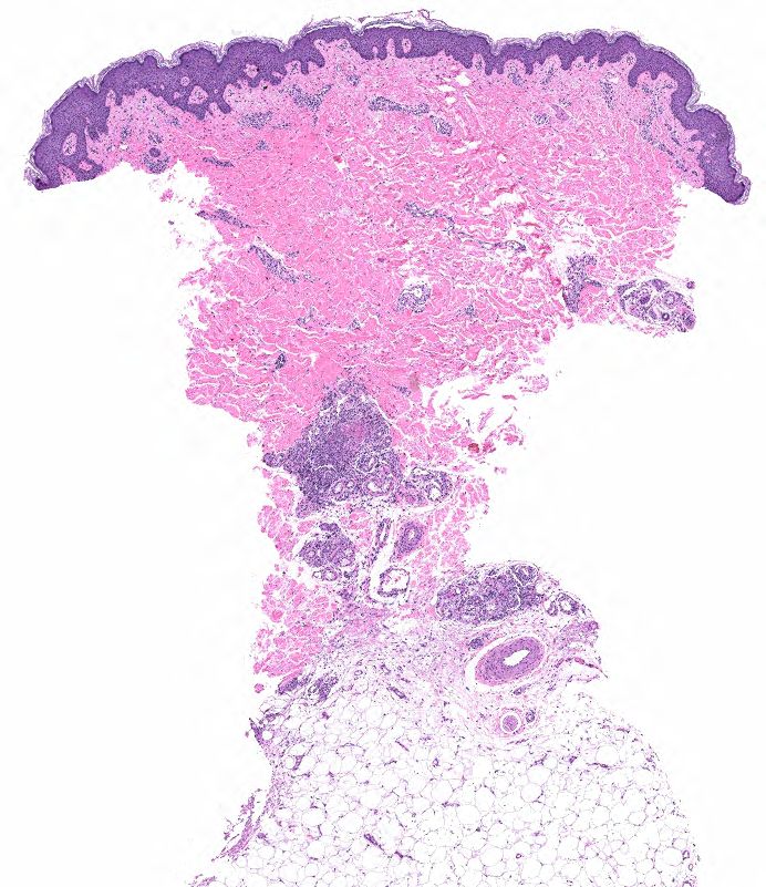
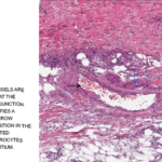
FIGURE 2A: Punch biopsy demonstrates an uninvolved epidermis with focal mid to deep dermal vasculocentric inflammation (H&E stain, 2x). (Click to enlarge.)

Rheumatoid factor was mildly positive at 17 IU/mL (RR: <14 IU/mL); antinuclear antibody (ANA) was 1:80, dense and fine speckled (RR: <1:80); angiotensin-converting enzyme (ACE) was normal. ANCA, myeloperoxidase antibodies (MPO), proteinase 3 (PR3) and cryoglobulin tests were negative. C3 and C4 were normal. Urinalysis returned 1+ protein and 1+ ketones; urine protein/creatinine was 0.10 mg/1.0 mg (RR: <0.10 mg/1.0 mg). Serum protein electrophoresis was normal, and serum free

kappa/lambda was absent.

Chest X-ray and transthoracic echocardiogram were unrevealing.

Left knee joint aspiration yielded turbid, yellow synovial fluid, with 10,301 nucleated cells/μL (RR: ≥2,000 nucleated cells/μL is classified as inflammatory synovial fluid), less than 3,000 red blood cells and no crystals. Synovial fluid culture did not yield an organism.

Page: 1 2 3 4 5 | Single Page
Share: 

Filed under:ConditionsVasculitis Tagged with:case reportFellowsFellows Forumpolyarteritis nodosa

Related Articles

    Case Report: Diagnosing, Treating Hepatitis B-Linked Polyarteritis Nodosa

    September 17, 2019

    Hepatitis B virus (HBV) associated polyarteritis nodosa (PAN) is an increasingly rare vasculitis in developed countries due to advances in HBV vaccination and antiviral therapy. However, the condition does persist, and rheumatologists should consider it when evaluating vasculitis cases. Below, we discuss a case that illustrates the varied clinical presentations PAN can encompass. A high…

    Medium-sized blood vessels are seen in the dermis and at the dermal-subcutaneous junction. The black arrow identifies a venule, and the blue arrow points to a fibrin deposition in the vessel wall. Extravasated neutrophils and erythrocytes are seen in the interstitium.

    Case Report: A Polyarteritis Nodosa Conundrum

    March 14, 2022

    Polyarteritis nodosa (PAN) is a systemic necrotizing vasculitis that typically affects medium-sized muscular arteries. The clinical subsets of PAN are idiopathic, generalized, secondary hepatitis B virus (HBV) associated and cutaneous PAN. These clinical subsets are important because of their therapeutic implications. Virtually any organ system can be affected in generalized PAN, but this vasculitis tends…

    Vasculitis Guidelines in Focus, Part 4: Polyarteritis Nodosa

    September 22, 2021

    Jason Springer, MD, MS, served as one of the lead authors of the ACR/VF guidelines for polyarteritis nodosa (PAN), and talks about the recommendations specific to PAN here.

    Alisusha; Trueffelpix / shutterstock.com

    3 New Vasculitis Guidelines Discussed

    May 13, 2021

    The ACR, in concert with the Vasculitis Foundation, released three new vasculitis guidelines online first in July, with a fourth slated for publication by the end of the year. These are the first ever to be produced and endorsed by the ACR and the Vasculitis Foundation. Although most of the recommendations are conditional, due to…

  • About Us
  • Meet the Editors
  • Issue Archives
  • Contribute
  • Advertise
  • Contact Us
  • Copyright © 2025 by John Wiley & Sons, Inc. All rights reserved, including rights for text and data mining and training of artificial technologies or similar technologies. ISSN 1931-3268 (print). ISSN 1931-3209 (online).
  • DEI Statement
  • Privacy Policy
  • Terms of Use
  • Cookie Preferences