Video: Every Case Tells a Story| Webinar: ACR/CHEST ILD Guidelines in Practice

An official publication of the ACR and the ARP serving rheumatologists and rheumatology professionals

  • Conditions
    • Axial Spondyloarthritis
    • Gout and Crystalline Arthritis
    • Myositis
    • Osteoarthritis and Bone Disorders
    • Pain Syndromes
    • Pediatric Conditions
    • Psoriatic Arthritis
    • Rheumatoid Arthritis
    • Sjögren’s Disease
    • Systemic Lupus Erythematosus
    • Systemic Sclerosis
    • Vasculitis
    • Other Rheumatic Conditions
  • FocusRheum
    • ANCA-Associated Vasculitis
    • Axial Spondyloarthritis
    • Gout
    • Psoriatic Arthritis
    • Rheumatoid Arthritis
    • Systemic Lupus Erythematosus
  • Guidance
    • Clinical Criteria/Guidelines
    • Ethics
    • Legal Updates
    • Legislation & Advocacy
    • Meeting Reports
      • ACR Convergence
      • Other ACR meetings
      • EULAR/Other
    • Research Rheum
  • Drug Updates
    • Analgesics
    • Biologics/DMARDs
  • Practice Support
    • Billing/Coding
    • EMRs
    • Facility
    • Insurance
    • QA/QI
    • Technology
    • Workforce
  • Opinion
    • Patient Perspective
    • Profiles
    • Rheuminations
      • Video
    • Speak Out Rheum
  • Career
    • ACR ExamRheum
    • Awards
    • Career Development
  • ACR
    • ACR Home
    • ACR Convergence
    • ACR Guidelines
    • Journals
      • ACR Open Rheumatology
      • Arthritis & Rheumatology
      • Arthritis Care & Research
    • From the College
    • Events/CME
    • President’s Perspective
  • Search

A Case of Nodular Rash & Painful Joints

Vania Lin, MD, MPH, Rebecca Johnson, MD, & Lisa Suter, MD  |  Issue: October 2022  |  October 10, 2022

Given the rarity of cutaneous PAN and its predominant distribution among individuals in their 40s or 50s, we considered a wide differential diagnosis for this patient when carrying out our evaluation, including infectious (e.g., disseminated gonococcal infection, syphilis, parvovirus B19 infection, Lyme disease, Rocky Mountain spotted fever, infective endocarditis, septic arthritis, tuberculosis), post-infectious (e.g., reactive arthritis, acute rheumatic fever) and rheumatological (e.g., ANCA-associated vasculitis, cryoglobulinemia, polyarteritis nodosa, sarcoidosis, rheumatoid arthritis, spondyloarthritis, systemic lupus erythematosus, crystalline arthritis) etiologies.5

He was ultimately diagnosed with cutaneous PAN via skin biopsy, with high-titer ASO suggestive of antecedent group A Streptococcal infection and possible subsequent rheumatic fever, and his treatment was tailored to this diagnosis. He had no apparent deep organ involvement to suggest a systemic vasculitis. Continued monitoring and heightened awareness are important moving forward, given the often chronic and relapsing course of cutaneous PAN and possible progression to systemic PAN.

ad goes here:advert-1
ADVERTISEMENT
SCROLL TO CONTINUE

Dr. Lin

Dr. Vania Lin

Vania Lin, MD, MPH, is a rheumatology fellow at Yale School of Medicine, New Haven, Conn.

Dr. Rebecca Johnson

Dr. Rebecca Johnson

Rebecca Johnson, MD, recently completed the Dermatopathology Fellowship Program at Yale School of Medicine, New Haven, Conn.

Dr. Suter

Dr. Suter

Lisa Suter, MD, is a professor of medicine in the Section of Rheumatology at the Yale School of Medicine, New Haven, Conn.; she is also director of quality measurement programs at the Yale New Haven Health Services Corporation Center for Outcomes Research and Evaluation (CORE).

Disclosures

Outside the submitted work, Dr. Suter receives support for directing a federal contract, the Measure & Instrument Development Support (MIDS) contract; Development, Reevaluation and Implementation of Outcome/Efficiency Measures for Hospital and Eligible Clinicians, funded by the CMS; and during the conduct of the study, grants from Brigham and Women’s Hospital (BWH). Dr. Suter received $5,000 or less per year in consulting fees to Dr. Losina, PI, on an NIH grant through BWH to study knee osteoarthritis.

References

  1. Haugeberg G, Bie R, Bendvold A, et al. Primary vasculitis in a Norwegian community hospital: A retrospective study. Clin Rheumatol. 1998;17(5):364–368.
  2. Reinhold-Keller E, Zeidler A, Gutfleisch J, et al. Giant cell arteritis is more prevalent in urban than in rural populations: Results of an epidemiological study of primary systemic vasculitides in Germany. Rheumatology (Oxford). 2000 Dec;39(12):1396–1402.
  3. Mahr A, Guillevin L, Poissonnet M, Aymé S. Prevalences of polyarteritis nodosa, microscopic polyangiitis, Wegener’s granulomatosis, and Churg-Strauss syndromein a French urban multiethnic population in 2000: A capture-recapture estimate. Arthritis Rheum. 2004 Feb 15;51(1):92–99.
  4. Pagnoux C, Seror R, Henegar C, et al. Clinical features and outcomes in 348 patients with polyarteritis nodosa: A systematic retrospective study of patients diagnosed between 1963 and 2005 and entered into the French Vasculitis Study Group Database. Arthritis Rheum. 2010 Feb;62(2):616–626.
  5. Daoud MS, Hutton KP, Gibson LE. Cutaneous periarteritis nodosa: A clinicopathological study of 79 cases. Br J Dermatol. 1997 May;136(5):706–713.
  6. Criado PR, Marques GF, Morita TCAB, de Carvalho JF. Epidemiological, clinical and laboratory profiles of cutaneous polyarteritis nodosa patients: Report of 22 cases and literature review. Autoimmun Rev. 2016 Jun;15(6):558–563.

Page: 1 2 3 4 5 | Single Page
Share: 

Filed under:ConditionsVasculitis Tagged with:case reportFellowsFellows Forumpolyarteritis nodosa

Related Articles

    Case Report: Diagnosing, Treating Hepatitis B-Linked Polyarteritis Nodosa

    September 17, 2019

    Hepatitis B virus (HBV) associated polyarteritis nodosa (PAN) is an increasingly rare vasculitis in developed countries due to advances in HBV vaccination and antiviral therapy. However, the condition does persist, and rheumatologists should consider it when evaluating vasculitis cases. Below, we discuss a case that illustrates the varied clinical presentations PAN can encompass. A high…

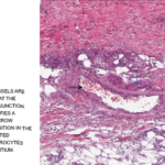
    Medium-sized blood vessels are seen in the dermis and at the dermal-subcutaneous junction. The black arrow identifies a venule, and the blue arrow points to a fibrin deposition in the vessel wall. Extravasated neutrophils and erythrocytes are seen in the interstitium.

    Case Report: A Polyarteritis Nodosa Conundrum

    March 14, 2022

    Polyarteritis nodosa (PAN) is a systemic necrotizing vasculitis that typically affects medium-sized muscular arteries. The clinical subsets of PAN are idiopathic, generalized, secondary hepatitis B virus (HBV) associated and cutaneous PAN. These clinical subsets are important because of their therapeutic implications. Virtually any organ system can be affected in generalized PAN, but this vasculitis tends…

    Vasculitis Guidelines in Focus, Part 4: Polyarteritis Nodosa

    September 22, 2021

    Jason Springer, MD, MS, served as one of the lead authors of the ACR/VF guidelines for polyarteritis nodosa (PAN), and talks about the recommendations specific to PAN here.

    Alisusha; Trueffelpix / shutterstock.com

    3 New Vasculitis Guidelines Discussed

    May 13, 2021

    The ACR, in concert with the Vasculitis Foundation, released three new vasculitis guidelines online first in July, with a fourth slated for publication by the end of the year. These are the first ever to be produced and endorsed by the ACR and the Vasculitis Foundation. Although most of the recommendations are conditional, due to…

  • About Us
  • Meet the Editors
  • Issue Archives
  • Contribute
  • Advertise
  • Contact Us
  • Copyright © 2025 by John Wiley & Sons, Inc. All rights reserved, including rights for text and data mining and training of artificial technologies or similar technologies. ISSN 1931-3268 (print). ISSN 1931-3209 (online).
  • DEI Statement
  • Privacy Policy
  • Terms of Use
  • Cookie Preferences