Video: Every Case Tells a Story| Webinar: ACR/CHEST ILD Guidelines in Practice

An official publication of the ACR and the ARP serving rheumatologists and rheumatology professionals

  • Conditions
    • Axial Spondyloarthritis
    • Gout and Crystalline Arthritis
    • Myositis
    • Osteoarthritis and Bone Disorders
    • Pain Syndromes
    • Pediatric Conditions
    • Psoriatic Arthritis
    • Rheumatoid Arthritis
    • Sjögren’s Disease
    • Systemic Lupus Erythematosus
    • Systemic Sclerosis
    • Vasculitis
    • Other Rheumatic Conditions
  • FocusRheum
    • ANCA-Associated Vasculitis
    • Axial Spondyloarthritis
    • Gout
    • Psoriatic Arthritis
    • Rheumatoid Arthritis
    • Systemic Lupus Erythematosus
  • Guidance
    • Clinical Criteria/Guidelines
    • Ethics
    • Legal Updates
    • Legislation & Advocacy
    • Meeting Reports
      • ACR Convergence
      • Other ACR meetings
      • EULAR/Other
    • Research Rheum
  • Drug Updates
    • Analgesics
    • Biologics/DMARDs
  • Practice Support
    • Billing/Coding
    • EMRs
    • Facility
    • Insurance
    • QA/QI
    • Technology
    • Workforce
  • Opinion
    • Patient Perspective
    • Profiles
    • Rheuminations
      • Video
    • Speak Out Rheum
  • Career
    • ACR ExamRheum
    • Awards
    • Career Development
  • ACR
    • ACR Home
    • ACR Convergence
    • ACR Guidelines
    • Journals
      • ACR Open Rheumatology
      • Arthritis & Rheumatology
      • Arthritis Care & Research
    • From the College
    • Events/CME
    • President’s Perspective
  • Search

A Primer on Imaging in Myositis

Rochelle Castillo, MD, MS, Andro Licaros, MD, & Jemima Albayda, MD  |  Issue: November 2022  |  November 9, 2022

Even after the diagnosis of IIM has been established, MRI remains useful to monitor response to treatment, qualitatively measure disease activity, and direct future management. This is particularly important when discordance exists between the patients’ symptoms and providers’ assessments and when routine serologic markers of muscle breakdown fail to correlate with clinical findings. Active disease requiring escalation or modification of therapy is indicated by more pronounced muscle edema, whereas response to therapy appears as resolution or reduction of edema (normalization of intensity values or “return to isointensity” in the above sequences). Notably, a relationship between the degree of STIR hyper­intensity and the abundance of pre-treatment inflammatory infiltrates on muscle biopsy has been shown in several studies.4,6

On the other hand, intensification of the largely irreversible markers of chronicity and damage, such as fatty replacement and atrophy (increased signal intensity and decreased muscle mass, respectively, on T1W), warrant reevaluation of the drug regimen to balance out the need to prevent both disease progression and unnecessary exposure to potentially harmful medications. Quantitative MRI studies have similarly demonstrated a modest correlation between the extent of fatty infiltration noted on MRI and that seen on muscle histopathology.7,8

ad goes here:advert-1
ADVERTISEMENT
SCROLL TO CONTINUE

Thus, the capacity of MRI findings to correlate with response to therapy underscores its value in guiding subsequent decision making. With its versatility, granularity and increasing ubiquity, it’s easy to see why MRI is the go-to muscle imaging modality (see Table 2).

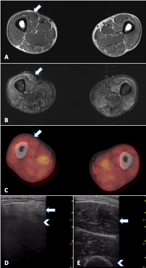
FIGURE 1. MRI, PET/CT and ultrasound in a 33-year-old man with refractory PM-Scl dermatomyositis. Arrows: rectus femoris; arrowheads: humerus. A) MRI of the thighs (T1W) showing no significant fatty replacement. B) MRI of thighs (STIR) with mild to moderate muscle edema (increased signal) mainly in anterior compartment. C) PET/CT showing symmetric bilateral increased FDG uptake in scattered muscles, especially pelvic adductors, D) increased muscle echointensity in the rectus femoris with attenuation (loss of bone echo of underlying femur) and atrophy in comparison with E) normal rectus femoris in age-matched control.

Ultrasound

Ultrasound is undeniable proof that oldies can still be goodies. With the advent of higher frequency probes, imaging resolution for soft tissue is now far higher than MRI and offers a close-up view of the muscle, which has its own advantages. Ultrasound also has more widespread availability, portability, cost efficiency, and real-time imaging capability, with the latter rendering it a useful tool for optimizing muscle biopsy site selection as well as for detecting abnormal muscle movements (see Table 2). However, ultrasound is heavily operator dependent, and knowledge of both proper image acquisition and image interpretation for muscle is needed.

ad goes here:advert-2
ADVERTISEMENT
SCROLL TO CONTINUE

Page: 1 2 3 4 5 | Single Page
Share: 

Filed under:ConditionsMyositis Tagged with:dermatomyositisMagnetic resonance imaging (MRI)myositispositron emission tomography (PET)Ultrasound

Related Articles

    Myositis Mysteries

    January 1, 2008

    Why isn’t my myositis patient getting better?

    New Tools for Myositis Diagnosis, Classification & Management

    April 15, 2019

    CHICAGO—At Hot Topics in Myositis, a session at the 2018 ACR/ARHP Annual Meeting, three experts discussed new classification criteria for idiopathic inflammatory myopathies (IIM) and offered practical primers on overlap myositis conditions and inclusion body myositis (IBM). New Myositis Classification Criteria After a 10-year development process, the new EULAR/ACR Classification Criteria for Adult and Juvenile…

    Autoantibodies in Autoimmune Myopathy

    Autoantibodies in Autoimmune Myopathy

    September 18, 2017

    In recent years, scientists and clinicians have learned a great deal about autoantibodies occurring in idiopathic inflammatory myopathies (IIMs). These new discoveries have reshaped our understanding of distinct clinical pheno­types in IIMs. Scientists continue to learn more about how these auto­antibodies shape pathophysiology, diagnosis, disease monitoring, prognosis and optimum treatment. Moving forward, these autoantibodies will…

    From Strength to Strength: Idiopathic Inflammatory Myopathy Diagnosis & Management

    December 2, 2021

    During the ACR Convergence 2021 Review Course, Rohit Aggarwal, MD, MS, provided an update on idiopathic inflammatory myopathy.

  • About Us
  • Meet the Editors
  • Issue Archives
  • Contribute
  • Advertise
  • Contact Us
  • Copyright © 2025 by John Wiley & Sons, Inc. All rights reserved, including rights for text and data mining and training of artificial technologies or similar technologies. ISSN 1931-3268 (print). ISSN 1931-3209 (online).
  • DEI Statement
  • Privacy Policy
  • Terms of Use
  • Cookie Preferences