Video: Every Case Tells a Story| Webinar: ACR/CHEST ILD Guidelines in Practice

An official publication of the ACR and the ARP serving rheumatologists and rheumatology professionals

  • Conditions
    • Axial Spondyloarthritis
    • Gout and Crystalline Arthritis
    • Myositis
    • Osteoarthritis and Bone Disorders
    • Pain Syndromes
    • Pediatric Conditions
    • Psoriatic Arthritis
    • Rheumatoid Arthritis
    • Sjögren’s Disease
    • Systemic Lupus Erythematosus
    • Systemic Sclerosis
    • Vasculitis
    • Other Rheumatic Conditions
  • FocusRheum
    • ANCA-Associated Vasculitis
    • Axial Spondyloarthritis
    • Gout
    • Psoriatic Arthritis
    • Rheumatoid Arthritis
    • Systemic Lupus Erythematosus
  • Guidance
    • Clinical Criteria/Guidelines
    • Ethics
    • Legal Updates
    • Legislation & Advocacy
    • Meeting Reports
      • ACR Convergence
      • Other ACR meetings
      • EULAR/Other
    • Research Rheum
  • Drug Updates
    • Analgesics
    • Biologics/DMARDs
  • Practice Support
    • Billing/Coding
    • EMRs
    • Facility
    • Insurance
    • QA/QI
    • Technology
    • Workforce
  • Opinion
    • Patient Perspective
    • Profiles
    • Rheuminations
      • Video
    • Speak Out Rheum
  • Career
    • ACR ExamRheum
    • Awards
    • Career Development
  • ACR
    • ACR Home
    • ACR Convergence
    • ACR Guidelines
    • Journals
      • ACR Open Rheumatology
      • Arthritis & Rheumatology
      • Arthritis Care & Research
    • From the College
    • Events/CME
    • President’s Perspective
  • Search

ACR Image Competition 2022 Results, Part 3

Masaki Itagane, MD  |  Issue: February 2023  |  February 15, 2023

Localized Scleroderma in Anti-NXP2-Antibody Positive Dermatomyositis

Click to enlarge.

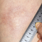
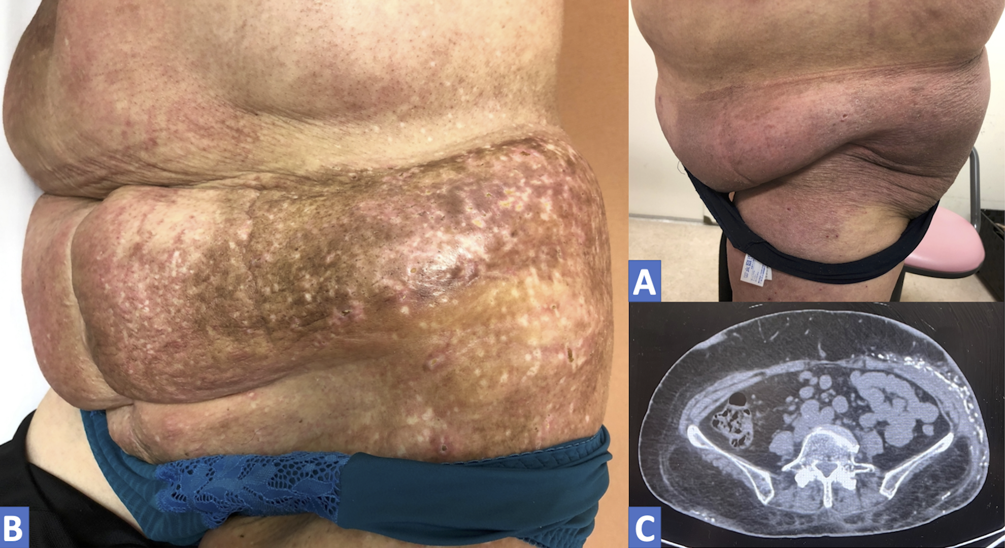
A 67-year-old woman presented with erythematous, indurated skin on her left flanks. She had been diagnosed with dermatomyositis one year earlier when proximal muscle weakness, dysphagia and skin rash developed (see Figure A). Tests at the time showed the presence of anti-NXP2 and anti-Ro52 antibodies, as well as pathological muscle findings. At that time, systemic glucocorticoids and mycophenolate mofetil alleviated her muscle symptoms, but the skin lesion progressively became hard, like stone, and discolored, with some depigmentation (see Figure B).

A skin biopsy conducted at the time of the current presentation showed vascular dilation and hyperplasia, inflammatory cell infiltrations and degenerated collagen fibers in subcutaneous tissue. A computed tomography scan of her abdomen showed calcinosis cutis (see Figure C). The localized scleroderma was not treated, but the patient is now doing well without worsening localized scleroderma.

ad goes here:advert-1
ADVERTISEMENT
SCROLL TO CONTINUE

Masaki Itagane, MD, is a rheumatologist in the Division of Rheumatology, Department of Internal Medicine at Okinawa Chubu Hospital, Okinawa, Japan.

About the Contest

The Rheumatology Image Library is a highly accessed teaching resource. The 2022 Image Competition sought images representing a diverse range of patients who show either characteristic or unusual manifestations of rheumatic disease, including systemic sclerosis, localized scleroderma and scleroderma mimics. Look for the Best Overall Image on our website and other regional winners in future issues or in the Rheumatology Image Library.

ad goes here:advert-2
ADVERTISEMENT
SCROLL TO CONTINUE

Share: 

Filed under:ConditionsMyositisSystemic Sclerosis Tagged with:calcinosisdermatomyositisImage Competition

Related Articles

    New Tools for Myositis Diagnosis, Classification & Management

    April 15, 2019

    CHICAGO—At Hot Topics in Myositis, a session at the 2018 ACR/ARHP Annual Meeting, three experts discussed new classification criteria for idiopathic inflammatory myopathies (IIM) and offered practical primers on overlap myositis conditions and inclusion body myositis (IBM). New Myositis Classification Criteria After a 10-year development process, the new EULAR/ACR Classification Criteria for Adult and Juvenile…

    Autoantibodies in Autoimmune Myopathy

    Autoantibodies in Autoimmune Myopathy

    September 18, 2017

    In recent years, scientists and clinicians have learned a great deal about autoantibodies occurring in idiopathic inflammatory myopathies (IIMs). These new discoveries have reshaped our understanding of distinct clinical pheno­types in IIMs. Scientists continue to learn more about how these auto­antibodies shape pathophysiology, diagnosis, disease monitoring, prognosis and optimum treatment. Moving forward, these autoantibodies will…

    Lost and found

    The History of ACE Inhibitors in Scleroderma Renal Crisis

    February 16, 2021

    Scleroderma renal crisis is a true medical emergency in rheumatology, one that requires prompt diagnosis and treatment. Here, we review the historic introduction of the angiotensin-converting enzyme inhibitors in this context, and highlight management and key questions moving forward. Background Awareness of renal disease in scleroderma dates back many years. The revered physician William Osler…

    Study Probes New Gene Therapy for Severe, Localized Scleroderma (Morphea)

    August 16, 2019

    In September 2018, the U.S. Food & Drug Administration (FDA) granted fast-track status to FCX‑013, a gene therapy product developed to treat moderate to severe localized scleroderma (morphea). Previously, the treatment received an orphan drug designation for localized scleroderma, as well as a rare pediatric disease designation. Phase 1 and 2 studies will assess safety…

  • About Us
  • Meet the Editors
  • Issue Archives
  • Contribute
  • Advertise
  • Contact Us
  • Copyright © 2025 by John Wiley & Sons, Inc. All rights reserved, including rights for text and data mining and training of artificial technologies or similar technologies. ISSN 1931-3268 (print). ISSN 1931-3209 (online).
  • DEI Statement
  • Privacy Policy
  • Terms of Use
  • Cookie Preferences