Video: Every Case Tells a Story| Webinar: ACR/CHEST ILD Guidelines in Practice

An official publication of the ACR and the ARP serving rheumatologists and rheumatology professionals

  • Conditions
    • Axial Spondyloarthritis
    • Gout and Crystalline Arthritis
    • Myositis
    • Osteoarthritis and Bone Disorders
    • Pain Syndromes
    • Pediatric Conditions
    • Psoriatic Arthritis
    • Rheumatoid Arthritis
    • Sjögren’s Disease
    • Systemic Lupus Erythematosus
    • Systemic Sclerosis
    • Vasculitis
    • Other Rheumatic Conditions
  • FocusRheum
    • ANCA-Associated Vasculitis
    • Axial Spondyloarthritis
    • Gout
    • Psoriatic Arthritis
    • Rheumatoid Arthritis
    • Systemic Lupus Erythematosus
  • Guidance
    • Clinical Criteria/Guidelines
    • Ethics
    • Legal Updates
    • Legislation & Advocacy
    • Meeting Reports
      • ACR Convergence
      • Other ACR meetings
      • EULAR/Other
    • Research Rheum
  • Drug Updates
    • Analgesics
    • Biologics/DMARDs
  • Practice Support
    • Billing/Coding
    • EMRs
    • Facility
    • Insurance
    • QA/QI
    • Technology
    • Workforce
  • Opinion
    • Patient Perspective
    • Profiles
    • Rheuminations
      • Video
    • Speak Out Rheum
  • Career
    • ACR ExamRheum
    • Awards
    • Career Development
  • ACR
    • ACR Home
    • ACR Convergence
    • ACR Guidelines
    • Journals
      • ACR Open Rheumatology
      • Arthritis & Rheumatology
      • Arthritis Care & Research
    • From the College
    • Events/CME
    • President’s Perspective
  • Search

ACR Image Competition 2024 Results, Part 5

Eaman Alhassan, MD  |  Issue: April 2025  |  April 5, 2025

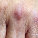
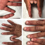
For the 2024 Image Competition, the ACR sought images with educational or remarkable manifestations representing a diverse range of pediatric patients with autoimmune, inflammatory, infectious and malignant drivers of rheumatic disease. Here, we showcase the winning images from North America.

Patient Presentation

Click to enlarge.

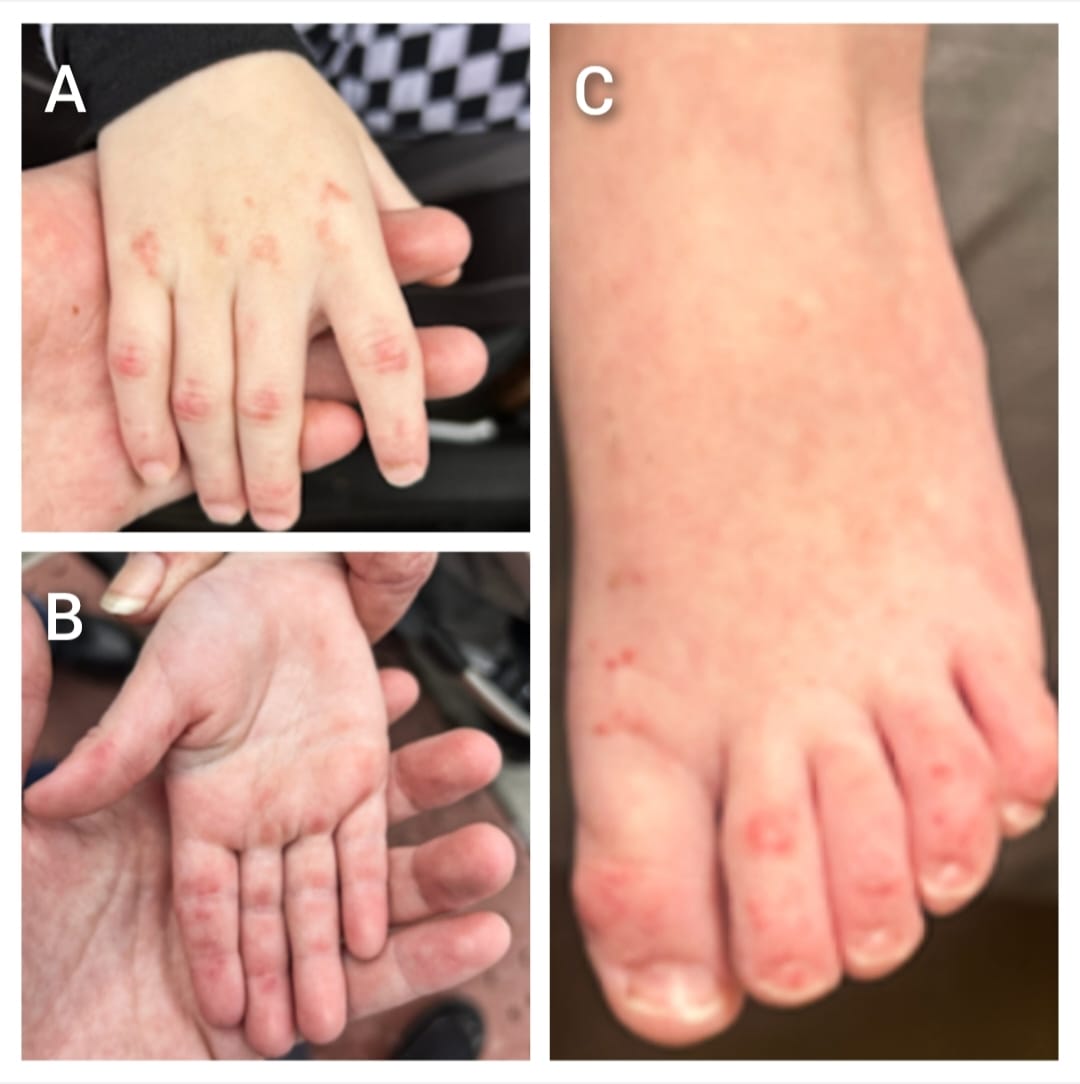
A 3-year-old boy presented with a four-month history of rash and hand blisters. On physical examination, the patient exhibited a heliotrope rash, Gottron papules (A), papules on the palmar creases of his fingers consistent with kissing papules (B), and erythematous desquamative plaques on the back of his toes (C).

ad goes here:advert-1
ADVERTISEMENT
SCROLL TO CONTINUE

Laboratory tests were positive for anti-melanoma differentiation-associated gene 5 (MDA-5) autoantibody, but normal muscle enzymes. Subtle myositis was evident on the magnetic resonance imaging (MRI) scan. High-resolution computed tomography (HRCT) of the chest was normal.

Kissing papules refer to papules occurring on the palmar creases of the fingers, forming symmetrical lesions on each side. It is one of the unique cutaneous characteristics of anti-MDA-5 dermatomyositis.

ad goes here:advert-2
ADVERTISEMENT
SCROLL TO CONTINUE

Learn More

For information about anti-MDA-5 dermatomyositis, read these articles from Arthritis & Rheumatology:

  • Understanding the Role of Type I Interferons in Cutaneous Lupus and Dermatomyositis: Toward Better Therapeutics: https://acrjournals.onlinelibrary.wiley.com/doi/10.1002/art.42983
  • Expert Perspective: Diagnostic Approach to Differentiating Juvenile Dermatomyositis from Muscular Dystrophy: https://acrjournals.onlinelibrary.wiley.com/doi/10.1002/art.43057

Eaman Alhassan, MD, is an inflammatory muscle disease (myositis) fellow at the University of Pittsburgh

Medical Center (UPMC). She earned her medical degree from King Saud University, Riyadh, Saudi Arabia,

completed her residency at the University of Maryland Medical Center, Baltimore, and completed a

rheumatology fellowship at the University of Southern California, Keck School of Medicine, Los Angeles.

About the Contest

The Rheumatology Image Library is a highly accessed teaching resource, and images from the ACR’s annual contest are added to it, helping keep it dynamic and current. Look for the Best Overall and regional winners in future issues and on our website.

Share: 

Filed under:ConditionsMyositis Tagged with:anti-MDA-5 antibody dermatomyositisdermatomyositisimage case reportImage CompetitionPediatric

Related Articles
    Cancer-Associated Myositis: A Case Report & Review of the Literature

    Cancer-Associated Myositis: A Case Report & Review of the Literature

    February 17, 2019

    Since it was first reported in 1916, a correlation between inflammatory myopathies and cancer has been noted in several studies. Population studies have confirmed this relationship, and the phrase cancer-associated myopathy has entered the vernacular. Over the past decade, research efforts have shifted toward revealing associations between autoantibodies and clinical phenotypes. One subset of auto-antigens…

    Tashatuvango / shutterstock.com

    Myositis-Specific Antibodies Identified

    January 16, 2020

    The idiopathic inflammatory myopathies (IIM) encompass eight categories: 1) dermatomyositis (DM) in adults, 2) juvenile dermatomyositis, 3) amyopathic DM, 4) cancer-associated DM, 5) polymyositis, 6) immune-mediated necrotizing myopathy, 7) inclusion body myositis, and 8) overlap myositis.1 These categories help classify the myopathies based on clinical and histologic features. The incidence of IIM is estimated at…

    Fellows’ Forum Case Report: Palmar Fasciitis & Polyarthritis Syndrome

    November 16, 2015

    Case report: A 78-year-old Caucasian female presented to our outpatient rheumatology clinic with pain in her bilateral shoulders, hands and knees that began suddenly one month earlier. She admitted to stiffness in her hands lasting several hours, and expressed an inability to extend her fingers. She denied fever, rashes, jaw claudication, headache or visual changes….

    The initial physical examination was significant for a nonblanching, papular rash along the palmar aspects of the hands and digits, periungual erythema, and edema and tenderness of the proximal and distal interphalangeal joints of the hands.

    Case Report: A Patient with Clinically Amyotrophic Dermatomyositis & Associated ILD & RA Overlap

    May 15, 2020

    Clinically amyotrophic dermatomyositis (CADM), a subset of dermatomyositis (DM), is a rare autoimmune disease characterized by typical DM cutaneous findings (e.g., heliotrope rash, Gottron papules, Gottron sign) without evidence of myositis.1 The incidence of DM and CADM is approximately 9.63 per 1 million people and 2.08 per 1 million people, respectively.2 The association with development…

  • About Us
  • Meet the Editors
  • Issue Archives
  • Contribute
  • Advertise
  • Contact Us
  • Copyright © 2025 by John Wiley & Sons, Inc. All rights reserved, including rights for text and data mining and training of artificial technologies or similar technologies. ISSN 1931-3268 (print). ISSN 1931-3209 (online).
  • DEI Statement
  • Privacy Policy
  • Terms of Use
  • Cookie Preferences