Video: Every Case Tells a Story| Webinar: ACR/CHEST ILD Guidelines in Practice

An official publication of the ACR and the ARP serving rheumatologists and rheumatology professionals

  • Conditions
    • Axial Spondyloarthritis
    • Gout and Crystalline Arthritis
    • Myositis
    • Osteoarthritis and Bone Disorders
    • Pain Syndromes
    • Pediatric Conditions
    • Psoriatic Arthritis
    • Rheumatoid Arthritis
    • Sjögren’s Disease
    • Systemic Lupus Erythematosus
    • Systemic Sclerosis
    • Vasculitis
    • Other Rheumatic Conditions
  • FocusRheum
    • ANCA-Associated Vasculitis
    • Axial Spondyloarthritis
    • Gout
    • Psoriatic Arthritis
    • Rheumatoid Arthritis
    • Systemic Lupus Erythematosus
  • Guidance
    • Clinical Criteria/Guidelines
    • Ethics
    • Legal Updates
    • Legislation & Advocacy
    • Meeting Reports
      • ACR Convergence
      • Other ACR meetings
      • EULAR/Other
    • Research Rheum
  • Drug Updates
    • Analgesics
    • Biologics/DMARDs
  • Practice Support
    • Billing/Coding
    • EMRs
    • Facility
    • Insurance
    • QA/QI
    • Technology
    • Workforce
  • Opinion
    • Patient Perspective
    • Profiles
    • Rheuminations
      • Video
    • Speak Out Rheum
  • Career
    • ACR ExamRheum
    • Awards
    • Career Development
  • ACR
    • ACR Home
    • ACR Convergence
    • ACR Guidelines
    • Journals
      • ACR Open Rheumatology
      • Arthritis & Rheumatology
      • Arthritis Care & Research
    • From the College
    • Events/CME
    • President’s Perspective
  • Search

Are Sacroiliac Joint MRIs Accurate in Routine Clinical Practice?

Anna E.F. Hadsbjerg, Simon Krabbe, Nora Vladimirova, Adrian Ciurea, Kristyna Bubova, Monika Gregová, Michael Nissen, Burkhard Moeller, Raphael Micheroli, Susanne Pedersen, Jakub Zavada, Ziga Snoj, Karlo Pintaric, Bjorn Gudbjornsson, Ziga Rotar, Iris Eshed, Iwona Sudol-Szopinska, Kasper Gosvig, Torsten Diekhoff, Robert Lambert, Manouk de hOoge, Helena V.G. Elmo, Merete Hetland, Lykke Oernbjerg & Mikkel Ostergaard  |  December 2, 2024

Abstract 0224 presented at ACR Convergence 2024

Background/Purpose

Magnetic resonance imaging (MRI) of the sacroiliac joints is widely used for diagnosing and monitoring patients with spondyloarthritis (SpA). However, local radiologists may be less familiar [than rheumatologists] with assessing MRIs in patients with spondyloarthritis potentially leading to misinterpretations and affecting the final diagnosis. We compared assessments of sacroiliac joint MRIs in patients with a diagnosis of axial spondyloarthritis (axSpA) or psoriatic arthritis (PsA) in local MRI reports from routine care in five European countries with re-reads by central experts to estimate the extent of over- or under-reporting of features and misclassification.

Methods

We included patients with a diagnosis of axSpA or PsA in a clinical patient registry from one of five European countries (DANBIO, Denmark; SCQM, Switzerland; ATTRA, Czech Republic; biorx.si, Slovenia; ICEBIO, Iceland; all participating in the EuroSpA Research Collaboration Network) with an available MRI of the sacroiliac joints and a corresponding local MRI report. MRIs were collected and read centrally by two experienced readers, blinded to clinical (except sex and age) and other imaging information. Readers registered whether the MRI was overall indicative of axSpA. In case of disagreement, MRIs were adjudicated by an experienced musculoskeletal radiologist (member of the ASAS/EULAR MRI group). Further, presence/absence of inflammatory and structural lesions (details in Table 1) were registered. Similar information was extracted from the local reports. Findings in local reports and central assessments were compared using central reads as reference standard.
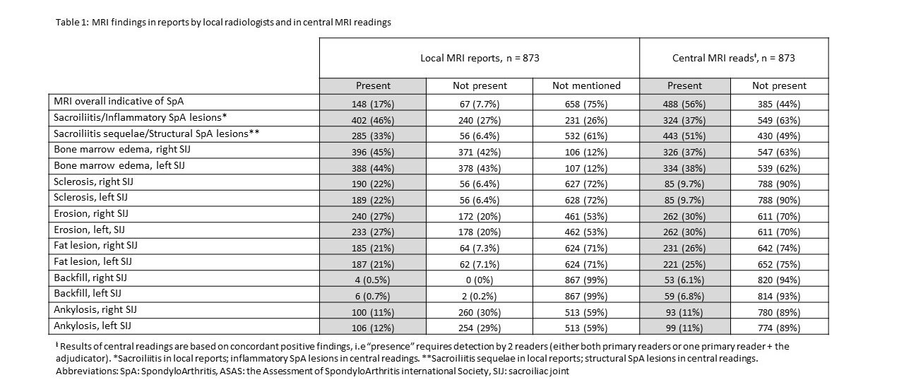
Table 1: MRI findings in reports by local radiologists and in central MRI readings.

ad goes here:advert-1
ADVERTISEMENT
SCROLL TO CONTINUE

Results

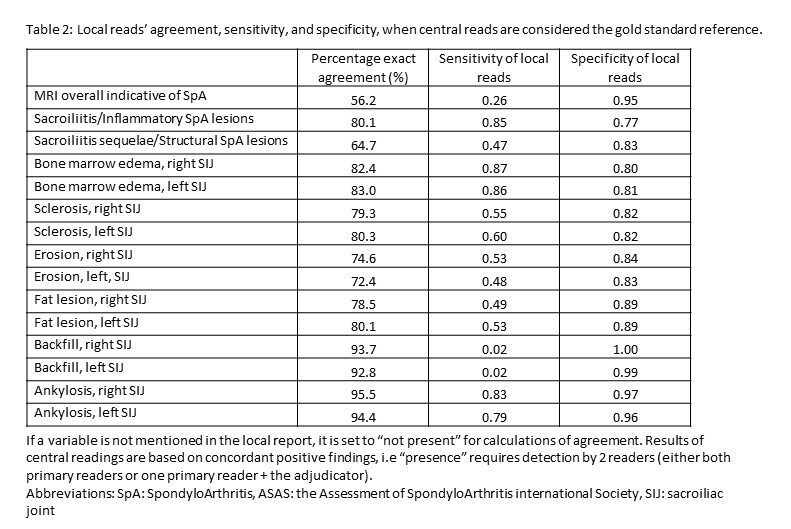
Overall, 873 patients with an MRI of the sacroiliac joints and a corresponding local MRI report were included; mean age 37.9 years, 475 (54%) male and 704/169 diagnosed with axSpA/PsA (see Table 1). Sacroiliitis/inflammatory SpA lesions were reported more often in local vs central reads (46% vs. 37%). Sacroiliitis sequelae/structural SpA lesions were reported less often in local vs. central reads (33% vs. 51%). In 17% of cases, local reads explicitly reported if the MRI was overall indicative of axSpA or not. On lesion level, bone marrow edema was the most frequent lesion judged present, both in local and central reads (right/left joint 45%/44% vs. 38%/37%, respectively), followed by erosion (27%/27% vs. 30%/30%). Compared with central reads, local reads tended to overestimate presence of bone marrow edema and sclerosis, underestimate/not assess backfill, while the frequency of erosions, fat and ankylosis was similar. Figure 1 shows examples with disagreement.Figure 1: Examples of cases with disagreement between local and central reads.
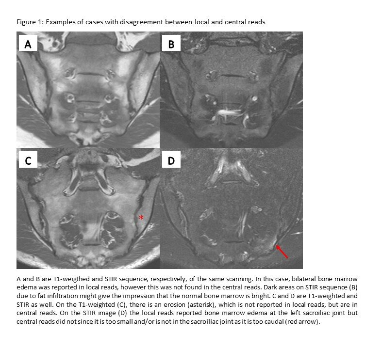
The sensitivity and specificity of local reads were 0.85 and 0.78 for sacroiliitis/inflammatory SpA lesions, and 0.48 and 0.83 for sacroiliitis sequelae/structural SpA lesions, respectively (see Table 2). For individual lesions, the sensitivity of local reads was highest for BME and ankylosis, followed by sclerosis. The specificity of local reads was ≥0.80 for all lesions.Table 2: Local reads' agreement, sensitivity, and specificity, when central reads are considered the gold standard reference.

Page: 1 2 3 | Single Page
Share: 

Filed under:ACR ConvergenceAmerican College of RheumatologyAxial SpondyloarthritisGuidelinesMeeting ReportsMeeting Reports Tagged with:ACR Convergence 2024ACR Convergence 2024 axSpA

Related Articles

    Imaging in Ankylosing Spondylitis

    April 1, 2015

    MRI inflammation, fat and new bone formation in the sacroiliac joints, spine in patients with AS

    Imaging of Axial Psoriatic Arthritis

    May 9, 2022

    The axial phenotype of psoriatic arthritis (axPsA) is an excellent example of a major controversy in rheumatology that has become the focus of attention because of the emergence of new therapies with different mechanisms of action for alleviating joint inflammation. It was first described in 1961 but, until recently, it has largely remained under the…

    Envision Arthritis Pathology

    April 1, 2008

    MRI advances in RA and OA

    The Art & Science of Imaging the Pediatric Sacroiliac Joint

    November 3, 2021

    Additional training may be needed on the appearance of the maturing sacroiliac (SI) joint on MRI. In a recent study, researchers found local radiologists may mistake normal physiologic changes of a maturing SI joint as sacroiliitis in pediatric patients.

  • About Us
  • Meet the Editors
  • Issue Archives
  • Contribute
  • Advertise
  • Contact Us
  • Copyright © 2025 by John Wiley & Sons, Inc. All rights reserved, including rights for text and data mining and training of artificial technologies or similar technologies. ISSN 1931-3268 (print). ISSN 1931-3209 (online).
  • DEI Statement
  • Privacy Policy
  • Terms of Use
  • Cookie Preferences