Video: Every Case Tells a Story| Webinar: ACR/CHEST ILD Guidelines in Practice

An official publication of the ACR and the ARP serving rheumatologists and rheumatology professionals

  • Conditions
    • Axial Spondyloarthritis
    • Gout and Crystalline Arthritis
    • Myositis
    • Osteoarthritis and Bone Disorders
    • Pain Syndromes
    • Pediatric Conditions
    • Psoriatic Arthritis
    • Rheumatoid Arthritis
    • Sjögren’s Disease
    • Systemic Lupus Erythematosus
    • Systemic Sclerosis
    • Vasculitis
    • Other Rheumatic Conditions
  • FocusRheum
    • ANCA-Associated Vasculitis
    • Axial Spondyloarthritis
    • Gout
    • Psoriatic Arthritis
    • Rheumatoid Arthritis
    • Systemic Lupus Erythematosus
  • Guidance
    • Clinical Criteria/Guidelines
    • Ethics
    • Legal Updates
    • Legislation & Advocacy
    • Meeting Reports
      • ACR Convergence
      • Other ACR meetings
      • EULAR/Other
    • Research Rheum
  • Drug Updates
    • Analgesics
    • Biologics/DMARDs
  • Practice Support
    • Billing/Coding
    • EMRs
    • Facility
    • Insurance
    • QA/QI
    • Technology
    • Workforce
  • Opinion
    • Patient Perspective
    • Profiles
    • Rheuminations
      • Video
    • Speak Out Rheum
  • Career
    • ACR ExamRheum
    • Awards
    • Career Development
  • ACR
    • ACR Home
    • ACR Convergence
    • ACR Guidelines
    • Journals
      • ACR Open Rheumatology
      • Arthritis & Rheumatology
      • Arthritis Care & Research
    • From the College
    • Events/CME
    • President’s Perspective
  • Search

Case Report: A Rare But Severe Complication of Dermatomyositis

Akrithi Udupa, MD, Paul McIntosh, MD, Thomas J. Cummings, MD, & Lisa Criscione-Schreiber, MD, Med  |  Issue: May 2021  |  May 13, 2021

Even with prompt recognition, acute hemorrhagic myositis can cause abrupt hemodynamic collapse & rapid progression to death.

His primary care physician consulted a hepatologist, due to concern for autoimmune hepatitis, and initiated empiric doxycycline with concern for tick-borne illness.

ad goes here:advert-1
ADVERTISEMENT
SCROLL TO CONTINUE

A recheck of laboratory studies during the current presentation revealed AST 191 U/L (normal: 15–41 U/L), ALT 76 U/L (normal: 17–63 U/L), ALP 112 U/L (normal: 24–110 U/L), albumin 1.8 g/dL (normal: 3.5–4.8 g/dL), aldolase 17.8 U/L (normal: <7.7 U/L), and peak creatine kinase (CK) 2,848 U/L (normal: 30–220 U/L). Creatinine and complete blood count tests were within normal limits. His CRP (2.79 mg/dL) and ferritin (735 ng/mL) were elevated, but his erythrocyte sedimentation rate was within the normal range.

Positive serologies included ANA 1:2560 with cytoplasmic speckling, AMA 39.9 U, RF 88 IU/mL, and anti-ribonuclear protein (RNP) 193 AU/mL. The MyoMarker Panel 3 was notable for an elevated anti-transcription intermediary factor 1-γ (TIF1-γ) at 126 U (normal: <20 U).

ad goes here:advert-2
ADVERTISEMENT
SCROLL TO CONTINUE

A contrast computerized tomography (CT) scan of his chest, abdomen and pelvis revealed numerous enlarged and heterogeneously enhancing mediastinal and right hilar lymph nodes (see Figure 2).

A contrasted CT scan of the chest shows numerous pathologic mediastinal and right hilar lymph nodes (maroon arrow).

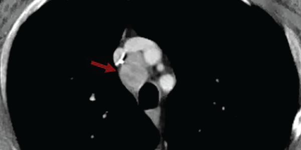
Figure 2. A contrasted CT scan of the chest shows numerous pathologic mediastinal and right hilar lymph nodes (maroon arrow).

Magnetic resonance imaging scan with myositis protocol of his lower extremities showed extensive symmetric T2 hyper­intense signal involving the bilateral thigh and pelvis musculature with evidence of subcutaneous and perifascial edema consistent with myositis (see Figure 3).

An MRI myositis protocol of the lower extremities with extensive, symmetric edema-like T2 signal of both thighs and the pelvis musculature.

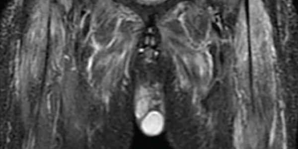
Figure 3. An MRI myositis protocol of the lower extremities with extensive, symmetric edema-like T2 signal of both thighs and the pelvis musculature.

Electromyography revealed abundant spontaneous activity in the form of fibrillation potentials and positive sharp waves in the deltoid and biceps brachii; it showed myopathic motor unit action potentials with early recruitment, consistent with an inflammatory myopathy.

A videofluoroscopic swallow study showed no oropharyngeal dysphagia. He underwent endobronchial ultrasound-guided fine needle aspiration biopsy of multiple lymph nodes. Rare tumor cells were present, with immunohistochemical testing positive for cytokeratin 7, suggestive of adenocarcinoma, and weakly positive for thyroid transcription factor 1, suggestive of a lung primary adenocarcinoma (see Figure 4).

An ultrasound-guided, fine-needle aspiration biopsy of the mediastinal lymph nodes shows rare tumor cells. Immunohistochemical testing revealed the presence of cytokeratin 7 and thyroid transcription factor 1.

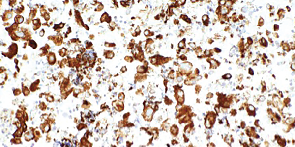
Figure 4. An ultrasound-guided, fine-needle aspiration biopsy of the mediastinal lymph nodes shows rare tumor cells. Immunohistochemical testing revealed the presence of cytokeratin 7 and thyroid transcription factor 1.

Next-generation sequencing was remarkable for missense mutations of NRAS, which are more commonly found in adenocarcinomas with a history of tobacco use, and TP53, which can be seen in a number of malignancies including lung cancers.2,3

Left forehead and right eyebrow lesion shave biopsies showed nodular and infiltrative pattern basal cell carcinoma, respectively. A right deltoid muscle biopsy demonstrated myopathic changes, fiber necrosis, fiber atrophy and minimal inflammatory cells, with no evidence of hemorrhage, granulomas or vasculitis (see Figure 5).

Page: 1 2 3 4 5 6 | Single Page
Share: 

Filed under:ConditionsMyositis Tagged with:case reportdermatomyositis (DM)hemorrhagic dermatomyositis

Related Articles

    New Tools for Myositis Diagnosis, Classification & Management

    April 15, 2019

    CHICAGO—At Hot Topics in Myositis, a session at the 2018 ACR/ARHP Annual Meeting, three experts discussed new classification criteria for idiopathic inflammatory myopathies (IIM) and offered practical primers on overlap myositis conditions and inclusion body myositis (IBM). New Myositis Classification Criteria After a 10-year development process, the new EULAR/ACR Classification Criteria for Adult and Juvenile…

    Autoantibodies in Autoimmune Myopathy

    Autoantibodies in Autoimmune Myopathy

    September 18, 2017

    In recent years, scientists and clinicians have learned a great deal about autoantibodies occurring in idiopathic inflammatory myopathies (IIMs). These new discoveries have reshaped our understanding of distinct clinical pheno­types in IIMs. Scientists continue to learn more about how these auto­antibodies shape pathophysiology, diagnosis, disease monitoring, prognosis and optimum treatment. Moving forward, these autoantibodies will…

    Tashatuvango / shutterstock.com

    Myositis-Specific Antibodies Identified

    January 16, 2020

    The idiopathic inflammatory myopathies (IIM) encompass eight categories: 1) dermatomyositis (DM) in adults, 2) juvenile dermatomyositis, 3) amyopathic DM, 4) cancer-associated DM, 5) polymyositis, 6) immune-mediated necrotizing myopathy, 7) inclusion body myositis, and 8) overlap myositis.1 These categories help classify the myopathies based on clinical and histologic features. The incidence of IIM is estimated at…

    Myositis Mysteries

    January 1, 2008

    Why isn’t my myositis patient getting better?

  • About Us
  • Meet the Editors
  • Issue Archives
  • Contribute
  • Advertise
  • Contact Us
  • Copyright © 2025 by John Wiley & Sons, Inc. All rights reserved, including rights for text and data mining and training of artificial technologies or similar technologies. ISSN 1931-3268 (print). ISSN 1931-3209 (online).
  • DEI Statement
  • Privacy Policy
  • Terms of Use
  • Cookie Preferences