Video: Every Case Tells a Story| Webinar: ACR/CHEST ILD Guidelines in Practice

An official publication of the ACR and the ARP serving rheumatologists and rheumatology professionals

  • Conditions
    • Axial Spondyloarthritis
    • Gout and Crystalline Arthritis
    • Myositis
    • Osteoarthritis and Bone Disorders
    • Pain Syndromes
    • Pediatric Conditions
    • Psoriatic Arthritis
    • Rheumatoid Arthritis
    • Sjögren’s Disease
    • Systemic Lupus Erythematosus
    • Systemic Sclerosis
    • Vasculitis
    • Other Rheumatic Conditions
  • FocusRheum
    • ANCA-Associated Vasculitis
    • Axial Spondyloarthritis
    • Gout
    • Psoriatic Arthritis
    • Rheumatoid Arthritis
    • Systemic Lupus Erythematosus
  • Guidance
    • Clinical Criteria/Guidelines
    • Ethics
    • Legal Updates
    • Legislation & Advocacy
    • Meeting Reports
      • ACR Convergence
      • Other ACR meetings
      • EULAR/Other
    • Research Rheum
  • Drug Updates
    • Analgesics
    • Biologics/DMARDs
  • Practice Support
    • Billing/Coding
    • EMRs
    • Facility
    • Insurance
    • QA/QI
    • Technology
    • Workforce
  • Opinion
    • Patient Perspective
    • Profiles
    • Rheuminations
      • Video
    • Speak Out Rheum
  • Career
    • ACR ExamRheum
    • Awards
    • Career Development
  • ACR
    • ACR Home
    • ACR Convergence
    • ACR Guidelines
    • Journals
      • ACR Open Rheumatology
      • Arthritis & Rheumatology
      • Arthritis Care & Research
    • From the College
    • Events/CME
    • President’s Perspective
  • Search

Case Report: A Rare But Severe Complication of Dermatomyositis

Akrithi Udupa, MD, Paul McIntosh, MD, Thomas J. Cummings, MD, & Lisa Criscione-Schreiber, MD, Med  |  Issue: May 2021  |  May 13, 2021

H&E staining of a muscle biopsy demonstrates myopathic changes, fiber necrosis and fiber atrophy, albeit a paucity of inflammatory cells.

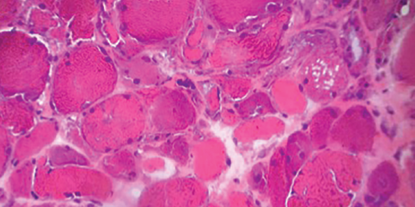
Figure 5. H&E staining of a muscle biopsy demonstrates myopathic changes, fiber necrosis and fiber atrophy, albeit a paucity of inflammatory cells.

MHC-I immunohistochemical staining showed patchy sarcolemmal up-regulation and an increased alkaline phosphatase staining pattern, consistent with an immune-mediated myopathic process. Electron microscopy of the muscle sample showed tubuloreticular inclusions (see Figure 6).

Electron microscopy of a muscle biopsy shows tubuloreticular inclusions (blue arrow).

Figure 6 . Electron microscopy of a muscle biopsy shows tubuloreticular inclusions (blue arrow).

Our patient was diagnosed with dermatomyositis and began therapy with 1 mg/kg of oral prednisone daily on hospital day (HD) 2.

ad goes here:advert-1
ADVERTISEMENT
SCROLL TO CONTINUE

On HD 5, he started to experience intermittent, deep, throbbing inguinal pain on the left side. Examination of the area revealed no cutaneous abnormalities, palpable mass, tenderness to palpation or significant pain with passive range of motion.

On HD 7, his hemoglobin dropped to 6.9 g/dL, and his blood pressure acutely dropped to 74/36 mmHg. Upon review of his laboratory studies, his CK had risen from 793 U/L on HD 5 to 1,100 U/L on HD 6 and to 1,729 U/L on HD 7. Prothrombin time and prothrombin INR were within normal range. Partial thromboplastin time was minimally elevated.

ad goes here:advert-2
ADVERTISEMENT
SCROLL TO CONTINUE

An ultrasound of his left groin showed a 6.9×2.3×4.4 cm anechoic collection most suspicious for a hematoma. A CT scan of his abdomen and pelvis with and without contrast revealed multiple intramuscular hematomas in the left psoas, left abductor magnus/pectineus and left rectus femoris.

His blood pressure normalized with blood products and intravenous fluids. With ongoing sensation of dysphagia and concern for severe features of dermatomyositis, treatment began with intravenous immunoglobulin (IVIG) 10% 0.5 g/kg per day on HD 9 for four days.

Notably, he had 10 doses of prophylactic subcutaneous heparin from HD 2 to HD 6. The hematomas were managed conservatively, and he recovered from the acute episode with supportive care. He received systemic chemotherapy for adenocarcinoma of presumed lung origin, started a slow oral prednisone steroid taper from 1 mg/kg per day, and was discharged to a long-term rehabilitation facility.

As an outpatient, he did not tolerate aggressive treatments with intravenous chemo­therapy, suffering from recurrent pulmonary infections that left him with an oxygen requirement and mostly bedbound. In view of this, he transitioned to a lower potency oral chemotherapeutic agent given his overall poor prognosis. Due to difficulty coordinating treatments and his ongoing health decline, he received no further IVIG. No additional hemorrhagic events were reported. Sadly, the patient died within a few months of discharge.

Page: 1 2 3 4 5 6 | Single Page
Share: 

Filed under:ConditionsMyositis Tagged with:case reportdermatomyositis (DM)hemorrhagic dermatomyositis

Related Articles

    New Tools for Myositis Diagnosis, Classification & Management

    April 15, 2019

    CHICAGO—At Hot Topics in Myositis, a session at the 2018 ACR/ARHP Annual Meeting, three experts discussed new classification criteria for idiopathic inflammatory myopathies (IIM) and offered practical primers on overlap myositis conditions and inclusion body myositis (IBM). New Myositis Classification Criteria After a 10-year development process, the new EULAR/ACR Classification Criteria for Adult and Juvenile…

    Autoantibodies in Autoimmune Myopathy

    Autoantibodies in Autoimmune Myopathy

    September 18, 2017

    In recent years, scientists and clinicians have learned a great deal about autoantibodies occurring in idiopathic inflammatory myopathies (IIMs). These new discoveries have reshaped our understanding of distinct clinical pheno­types in IIMs. Scientists continue to learn more about how these auto­antibodies shape pathophysiology, diagnosis, disease monitoring, prognosis and optimum treatment. Moving forward, these autoantibodies will…

    Tashatuvango / shutterstock.com

    Myositis-Specific Antibodies Identified

    January 16, 2020

    The idiopathic inflammatory myopathies (IIM) encompass eight categories: 1) dermatomyositis (DM) in adults, 2) juvenile dermatomyositis, 3) amyopathic DM, 4) cancer-associated DM, 5) polymyositis, 6) immune-mediated necrotizing myopathy, 7) inclusion body myositis, and 8) overlap myositis.1 These categories help classify the myopathies based on clinical and histologic features. The incidence of IIM is estimated at…

    Myositis Mysteries

    January 1, 2008

    Why isn’t my myositis patient getting better?

  • About Us
  • Meet the Editors
  • Issue Archives
  • Contribute
  • Advertise
  • Contact Us
  • Copyright © 2025 by John Wiley & Sons, Inc. All rights reserved, including rights for text and data mining and training of artificial technologies or similar technologies. ISSN 1931-3268 (print). ISSN 1931-3209 (online).
  • DEI Statement
  • Privacy Policy
  • Terms of Use
  • Cookie Preferences