Video: Every Case Tells a Story| Webinar: ACR/CHEST ILD Guidelines in Practice

An official publication of the ACR and the ARP serving rheumatologists and rheumatology professionals

  • Conditions
    • Axial Spondyloarthritis
    • Gout and Crystalline Arthritis
    • Myositis
    • Osteoarthritis and Bone Disorders
    • Pain Syndromes
    • Pediatric Conditions
    • Psoriatic Arthritis
    • Rheumatoid Arthritis
    • Sjögren’s Disease
    • Systemic Lupus Erythematosus
    • Systemic Sclerosis
    • Vasculitis
    • Other Rheumatic Conditions
  • FocusRheum
    • ANCA-Associated Vasculitis
    • Axial Spondyloarthritis
    • Gout
    • Psoriatic Arthritis
    • Rheumatoid Arthritis
    • Systemic Lupus Erythematosus
  • Guidance
    • Clinical Criteria/Guidelines
    • Ethics
    • Legal Updates
    • Legislation & Advocacy
    • Meeting Reports
      • ACR Convergence
      • Other ACR meetings
      • EULAR/Other
    • Research Rheum
  • Drug Updates
    • Analgesics
    • Biologics/DMARDs
  • Practice Support
    • Billing/Coding
    • EMRs
    • Facility
    • Insurance
    • QA/QI
    • Technology
    • Workforce
  • Opinion
    • Patient Perspective
    • Profiles
    • Rheuminations
      • Video
    • Speak Out Rheum
  • Career
    • ACR ExamRheum
    • Awards
    • Career Development
  • ACR
    • ACR Home
    • ACR Convergence
    • ACR Guidelines
    • Journals
      • ACR Open Rheumatology
      • Arthritis & Rheumatology
      • Arthritis Care & Research
    • From the College
    • Events/CME
    • President’s Perspective
  • Search

Case Report: Amyloid Beta-Related Angiitis of the Central Nervous System

Alexandros Grivas, MD, Nikil Revuri, MD, Aleksandra Karaseva, MD, Basimah AlBalooshy, MD, & Andres M. Ponce, MD  |  Issue: October 2024  |  October 8, 2024

A repeat brain MRI showed a 2.3 cm x 0.9 cm enhancing lesion in the right parasagittal frontal lobe, with extensive leptomeningeal involvement, multiple foci of hemorrhage within the regions of enhancement, and associated vasogenic edema (see Figure 1). MRI spectroscopy showed necrotic tissue, with findings less consistent with glial neoplasm, lymphoma or demyelinating disease (see Figure 2).

Due to inconclusive findings, an open right frontal lobe biopsy was performed. Preliminary results were consistent with necrotizing granulomatous vasculitis (i.e., necrotizing vasculitis including vessels with fibrinoid necrosis in a background of marked inflammation with CD68+ giant cells). No indications of lymphoma, glial neoplasm or microorganisms were found.

ad goes here:advert-1
ADVERTISEMENT
SCROLL TO CONTINUE

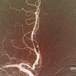
Differential diagnoses at that time included primary angiitis of the CNS, isolated CNS sarcoidosis and systemic granulomatous vasculitis.

A review of systems was unremarkable. Inflammatory markers were notable for a mildly elevated C-reactive protein (CPR; 1.4 mg/L) but normal erythrocyte sedimentation rate (ESR; 26 mm/hr), and an extensive autoimmune evaluation was negative. A temporal artery ultrasound was performed, showing no evidence of temporal arteritis. A review of the CT images also did not reveal any signs of systemic vasculitis or sarcoidosis.

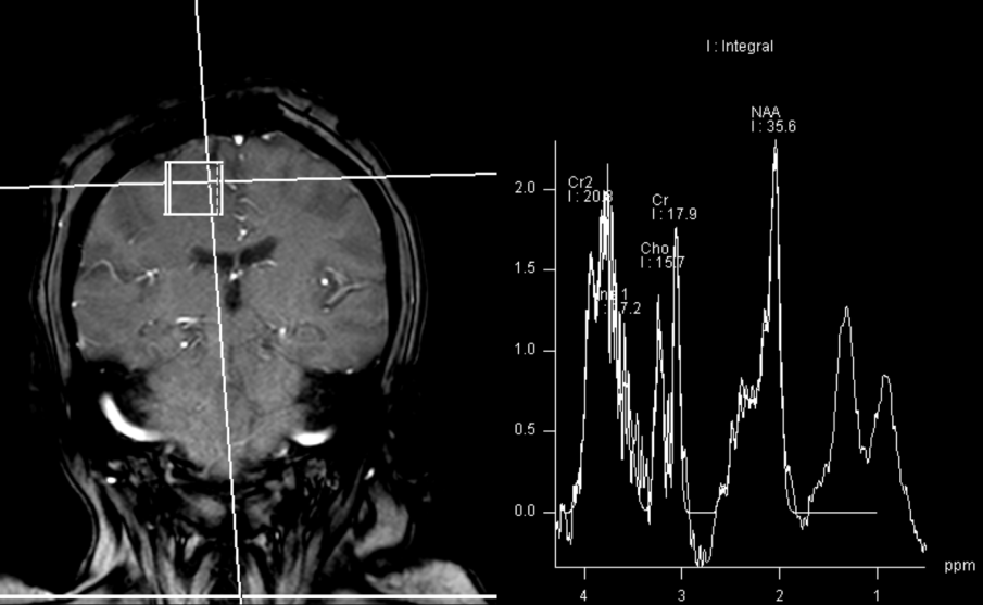
Figure 2: MRI spectroscopy demonstrated decreased NAA (N-acetylaspartate) peak without significant elevation of the choline peak. Findings are suggestive of a necrotic inflammatory process. Spectral features were inconsistent with glial neoplasm or other neoplastic etiologies, such as lymphoma or metastasis. (Click to enlarge.)

Given a low suspicion for infection, empiric treatment was started with pulse-dose glucocorticoids (1 g daily for three days), followed by 1 mg/kg/day of methylprednisolone. The patient exhibited rapid clinical response, with initial improvement in left upper arm strength within days of glucocorticoid initiation. The patient was discharged to a rehabilitation center and received six doses of cyclophosphamide as an outpatient, while remaining on low-dose prednisone (5 mg daily).

The final pathology report showed strong positive staining of the subarachnoid and parenchymal vessels with amyloid β, establishing the diagnosis of inflammatory cerebral amyloid angiopathy (CAA), specifically the cerebral amyloid β-related angiitis (ABRA) subtype (see Figure 3).

At a six-month follow-up in the rheumatology clinic, the patient demonstrated substantial clinical improvement, with complete resolution of her headaches, recovery of left upper extremity strength and improvement in left lower extremity weakness (4/5).

Discussion

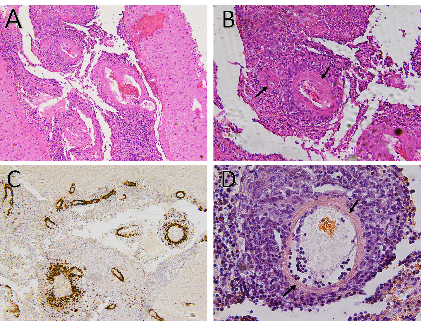
Figure 3A: Hematoxylin and eosin stain demonstrates small to medium-sized vessels that show necrotizing, fibrinoid necrosis representing a necrotizing vasculitis (100x).

Page: 1 2 3 4 5 6 | Single Page
Share: 

Filed under:ConditionsOther Rheumatic ConditionsVasculitis Tagged with:amyloid β-related angiitiscase reportcentral nervous system vasculitiscerebral amyloid angiopathy

Related Articles

    Meet the Challenge of Primary CNS Vasculitis

    September 1, 2011

    Diagnosis and treatment of this rare and poorly understood condition

    Case Report: Lymphocytic Vasculitis of the Central Nervous System

    January 17, 2019

    Vasculitis is a group of chronic inflammatory diseases in which the blood vessel is the target of an immune reaction. They can be secondary to connective tissue disease, idiopathic or due to infection, neoplasm or drugs.1 Primary angiitis of the central nervous system (PACNS) is a rare syndrome characterized by inflammatory cell infiltration and necrosis…

    The Legacy of Amyloid: Infiltration Linked to Dementia, Rheumatic Disease

    October 16, 2017

    An Unforgettable Story Her name was unforgettable. Not only did we share our given names—Simon and Simone, but her French-Canadian surname was based on this appellation, too. I was the junior resident working on our hospital’s nephrology service when she was admitted for evaluation of progressively worsening kidney disease and an overall failure to thrive….

    Rheumatology Case Report: When Moyamoya Disease Mimicks Primary Central Nervous System Vasculitis

    November 16, 2015

    Case report: A 60-year-old Hispanic male with poorly controlled hypertension was sent from the primary care clinic for evaluation of malignant hypertension with a systolic blood pressure above 200 mmHg. His symptoms at the time of presentation included episodic confusion, worsening vision and an unsteady gait. A head computed tomography (CT) scan showed a subacute…

  • About Us
  • Meet the Editors
  • Issue Archives
  • Contribute
  • Advertise
  • Contact Us
  • Copyright © 2025 by John Wiley & Sons, Inc. All rights reserved, including rights for text and data mining and training of artificial technologies or similar technologies. ISSN 1931-3268 (print). ISSN 1931-3209 (online).
  • DEI Statement
  • Privacy Policy
  • Terms of Use
  • Cookie Preferences