A 53-year-old man was hospitalized for pericarditis, abdominal lymphadenopathy of unknown origin and non-bloody diarrhea. He was admitted for four days, and then he was discharged home without incident.
Two months after his initial presentation, he was readmitted for the evaluation of several new issues, including symmetric arthralgias, hypovolemia with associated electrolyte abnormalities and concurrent weight loss. On arrival at the emergency department, his mental status was altered, but it improved after intravenous fluids were started and returned to baseline within 24 hours.
The arthralgias had started six weeks prior to his new admission. The joint pain had started in his left wrist. It then spread to the knees, ankles, hips and shoulders. The pain did not worsen with activity and was not relieved by rest. The patient reported he had noticed progressive hand swelling. His pain had worsened to the point that walking was unbearable. He reported fever, loss of appetite, nausea and vomiting. He also reported having had one or two extremely watery, mucous-laden stools just prior to his emergency department visit.
On admission, the patient was noted to have numerous laboratory abnormalities: hyponatremia with a sodium level of 130 mEq/L (reference range [RR]: 136–145); hypokalemia with potassium at 3.0 mEq/L (RR: 3.5–5.1); a chloride level of 92 mEq/L (RR: 98–107); hypocalcemia with a level of 7.9 mg/dL (RR: 8.5–10.1); aspartate aminotransferase of 109 units/L (RR: 15–37); alanine aminotransferase of 84 units/L (RR: 12–78); elevated alkaline phosphatase of 158 units/L (RR: 45–117); C-reactive protein of 19 mg/dL (RR: 0–0.3); an erythrocyte sedimentation rate (ESR) of 95 mm/hr (RR: 0–20); thyroid-stimulating hormone of 6.46 µIU/mL (RR: 0.36–3.74); albumin of 1.3 gm/dL (RR: 3.4–5.0); prothrombin time of 16.6 seconds (RR: 9.0–12.5); international normalized ratio of 1.6 (RR: 0.8–1.2); a white blood cell count of 21,900/µL (RR: 4,000–11,000), with an increase in the number of immature leukocytes; hemoglobin of 8.0 g/dL (RR: 12.0–18.0); hematocrit of 24.9% (RR: 36.0–55); and a platelet count of 445,000 µL (RR: 125,000–400,000), with a left shift noted on admission. His corrected calcium was 9.9 mg/dL (RR: 8.5–10.1).
Given the patient’s diarrhea, he underwent a polymerase chain reaction (PCR) test for Clostridium difficile, which was negative. Blood cultures and stool cultures did not reveal an infectious etiology. A rheumatologist was then consulted for evaluation of his symmetric inflammatory polyarthralgias with concurrent lymphadenopathy.
The patient denied any history of blood clots, seizure or stroke. He had no history of leukopenia, thrombocytopenia or anemia. He had no known family history of systemic lupus erythematosus, gout, psoriasis or rheumatoid arthritis. He did complain, however, of having dry eyes, dry mouth and pharyngitis.
The physical exam of the patient revealed a pink-tinged, pruritic, circumferential rash encompassing his neck and chest. He had abdominal distension with mild hepatosplenomegaly, but without evidence of ascites. We noted symmetric polyarthritis, involving both wrists, distal interphalangeal joints, proximal interphalangeal joints, metacarpophalangeal joints and his elbows. Palpable synovitis of the shoulders, knees and ankles was also noted. Trochanteric and subacromial bursitis was present bilaterally.
Tests for anti-nuclear antibodies (ANA), rheumatoid factor, cyclic citrullinated peptide (CCP) antibodies, double-stranded DNA, anti-Smith antibodies and anti-RNP antibodies were negative. C3 and C4 levels were within the normal range. Syphilis serology, hepatitis serology and HIV tests were all negative. Anti-streptolysin screening was negative as well. Creatine kinase levels were within the normal range; his myoglobin was 63 ng/mL (RR: 10–92), and his uric acid was 2.2 mg/dL (RR: 2.6–7.2). His ferritin was elevated at 1,906.8 ng/mL (RR: 8–388).
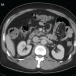
A computed tomography (CT) scan of the abdomen and pelvis was obtained to evaluate his lymphadenopathy, abdominal pain and diarrhea. The CT scan showed upper abdominal retroperitoneal lymphadenopathy, which was nonspecific in appearance and did not demonstrate evidence of necrosis or adjacent inflammatory changes (see Figure 1, above).
A diagnosis of adult-onset Still’s disease was suspected due to his clinical presentation and fulfillment of the Yamaguchi and Fautrel’s classification criteria.1,2 All major and minor criteria, with the exception of a fever, were satisfied. He had demonstrated persistent arthralgias for more than two weeks and experienced a sore throat, transient erythematous rash, leukocytosis with a left shift, lymphadenopathy, hepatosplenomegaly, elevated aminotransferases, negative rheumatoid factor and negative ANA (see Tables 1 & 2).