Video: Every Case Tells a Story| Webinar: ACR/CHEST ILD Guidelines in Practice

An official publication of the ACR and the ARP serving rheumatologists and rheumatology professionals

  • Conditions
    • Axial Spondyloarthritis
    • Gout and Crystalline Arthritis
    • Myositis
    • Osteoarthritis and Bone Disorders
    • Pain Syndromes
    • Pediatric Conditions
    • Psoriatic Arthritis
    • Rheumatoid Arthritis
    • Sjögren’s Disease
    • Systemic Lupus Erythematosus
    • Systemic Sclerosis
    • Vasculitis
    • Other Rheumatic Conditions
  • FocusRheum
    • ANCA-Associated Vasculitis
    • Axial Spondyloarthritis
    • Gout
    • Psoriatic Arthritis
    • Rheumatoid Arthritis
    • Systemic Lupus Erythematosus
  • Guidance
    • Clinical Criteria/Guidelines
    • Ethics
    • Legal Updates
    • Legislation & Advocacy
    • Meeting Reports
      • ACR Convergence
      • Other ACR meetings
      • EULAR/Other
    • Research Rheum
  • Drug Updates
    • Analgesics
    • Biologics/DMARDs
  • Practice Support
    • Billing/Coding
    • EMRs
    • Facility
    • Insurance
    • QA/QI
    • Technology
    • Workforce
  • Opinion
    • Patient Perspective
    • Profiles
    • Rheuminations
      • Video
    • Speak Out Rheum
  • Career
    • ACR ExamRheum
    • Awards
    • Career Development
  • ACR
    • ACR Home
    • ACR Convergence
    • ACR Guidelines
    • Journals
      • ACR Open Rheumatology
      • Arthritis & Rheumatology
      • Arthritis Care & Research
    • From the College
    • Events/CME
    • President’s Perspective
  • Search

Case Report: Contagious Rash in Active Systemic Lupus Erythematosus

Kenneth W. Van Dyke, DO, Jen K. Erbil, MD, and Harmanjot K. Grewal, MD  |  March 1, 2015

Figure 2: Anterior (a) and lateral (b) views of scalp showing nodular hyperkeratotic lesions with discoloration of involved skin.

Figure 2: Anterior (a) and lateral (b) views of scalp showing nodular hyperkeratotic lesions with discoloration of involved skin.

Diagnosis

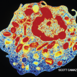
Crusted scabies, formerly known as Norwegian scabies, is an uncommon parasitic infection caused by infestation of the superficial layers of the skin with the mite, Sarcoptes scabiei var hominis.1 The name Norwegian scabies originates from its first description in 1848 of a group of Norwegian leprosy patients who developed the crusted scabies variant.2

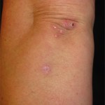
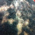
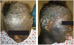
Unlike typical scabies, crusted scabies requires an inadequate host response to the arthropod mite, leading to a hyperinfestation state, with up to 2 million mites per person.3,4 This presentation usually occurs in patients with compromised immune systems, such as those on immunosuppressive therapy, infections with HIV or HTLV-1, or those with severe debilitation or cognitive dysfunction.1,5 Pruritis, caused by an allergic response to the mites within the skin, is a common manifestation of infection, which can lead to the diagnosis. However, in the immunocompromised patient, this urge to itch may be absent due to an altered immune response, which allows for heavy infestation.3,4

ad goes here:advert-2
ADVERTISEMENT
SCROLL TO CONTINUE

Crusted scabies is highly contagious and can be spread by minimal contact or indirect transmission through clothing or bedding. Classic scabies usually requires prolonged person-to-person contact and is rarely spread without direct contact.1 A classic infection of scabies has a mite load of about 10–15 mites per person; however, in crusted scabies, there are millions of mites underneath the skin, causing a crusted, scaly or even warty hyperkeratotic appearance of the skin.1,3

The presentation of crusted scabies is commonly limited to an isolated region of the body, such as the palms and soles, but infrequently involves the scalp. This atypical presentation can mimic many other conditions (e.g., dermatomyositis, psoriasis, discoid lupus) resulting in a delay in diagnosis.5 Prompt recognition of the condition is necessary because an outbreak can spread to those working close to the infected individual or even progress to an institution-wide infestation, as reported in our case.3 There is also heightened risk for secondary infections that could potentially lead to a life-threatening sepsis.2

The diagnosis of crusted scabies requires direct visualization of the mite with a light microscope under low power; however, false negatives can occur.

The diagnosis of crusted scabies requires direct visualization of the mite with a light microscope under low power; however, false negatives can occur. A mite may be visualized on a skin biopsy as well.1 Treatment for crusted scabies includes topical keratolytic therapy, topical scabicides and a multidose regimen of ivermectin depending on the severity of the condition.3,6

ad goes here:advert-1
ADVERTISEMENT
SCROLL TO CONTINUE

Page: 1 2 3 | Single Page
Share: 

Filed under:ConditionsResearch RheumSystemic Lupus Erythematosus Tagged with:DiagnosisrashrheumatologyscabiesSystemic lupus erythematosus

Related Articles
    Case Report: Skin-Deep Eosinophils

    Case Report: Skin-Deep Eosinophils

    May 17, 2018

    Eosinophilia is usually defined as an eosinophil count of more than 500/microL in peripheral blood.1 An eosinophil count of more than 1,500 is referred to as hypereosinophilia (HE); hypereosinophilic syndrome (HES) is defined as HE associated with organ dysfunction attributable to eosinophilia.2 Eosinophilia can occur due to infectious, malignancy, autoimmune or allergic etiologies. However, a…

    Case Study in Dermatology: Tender Papules on Elbows, Hands in RA

    February 17, 2016

    The Case A 41-year-old woman was referred to the dermatology clinic for a three-month history of tender lesions on her elbows and around the joints of her hands. Her medical history was notable for seropositive, non-erosive rheumatoid arthritis (RA), which was being treated with 5 mg pred­nisone daily, 10 mg methotrexate weekly by mouth, 200…

    Case Report: A Patient Presents with Rare, Fulminant SAPHO Syndrome

    October 18, 2018

    Synovitis, acne, pustulosis, hyperostosis and osteitis (SAPHO) syndrome is a heterogeneous, inflammatory, musculoskeletal disease. The disease is an insidious, sterile osteitis with associated skin and synovial inflammation.1 Diagnosis can prove challenging, but a thorough clinical history, high clinical suspicion and imaging techniques can help clinch it. The below case reveals a rare, fulminant presentation of…

    T Cells in Systemic Lupus Erythematosus

    August 1, 2011

    Progress toward targeted therapy

  • About Us
  • Meet the Editors
  • Issue Archives
  • Contribute
  • Advertise
  • Contact Us
  • Copyright © 2025 by John Wiley & Sons, Inc. All rights reserved, including rights for text and data mining and training of artificial technologies or similar technologies. ISSN 1931-3268 (print). ISSN 1931-3209 (online).
  • DEI Statement
  • Privacy Policy
  • Terms of Use
  • Cookie Preferences