At a follow-up with the rheumatologist, the patient’s scars were noted to have dramatically decreased in size.
Discussion
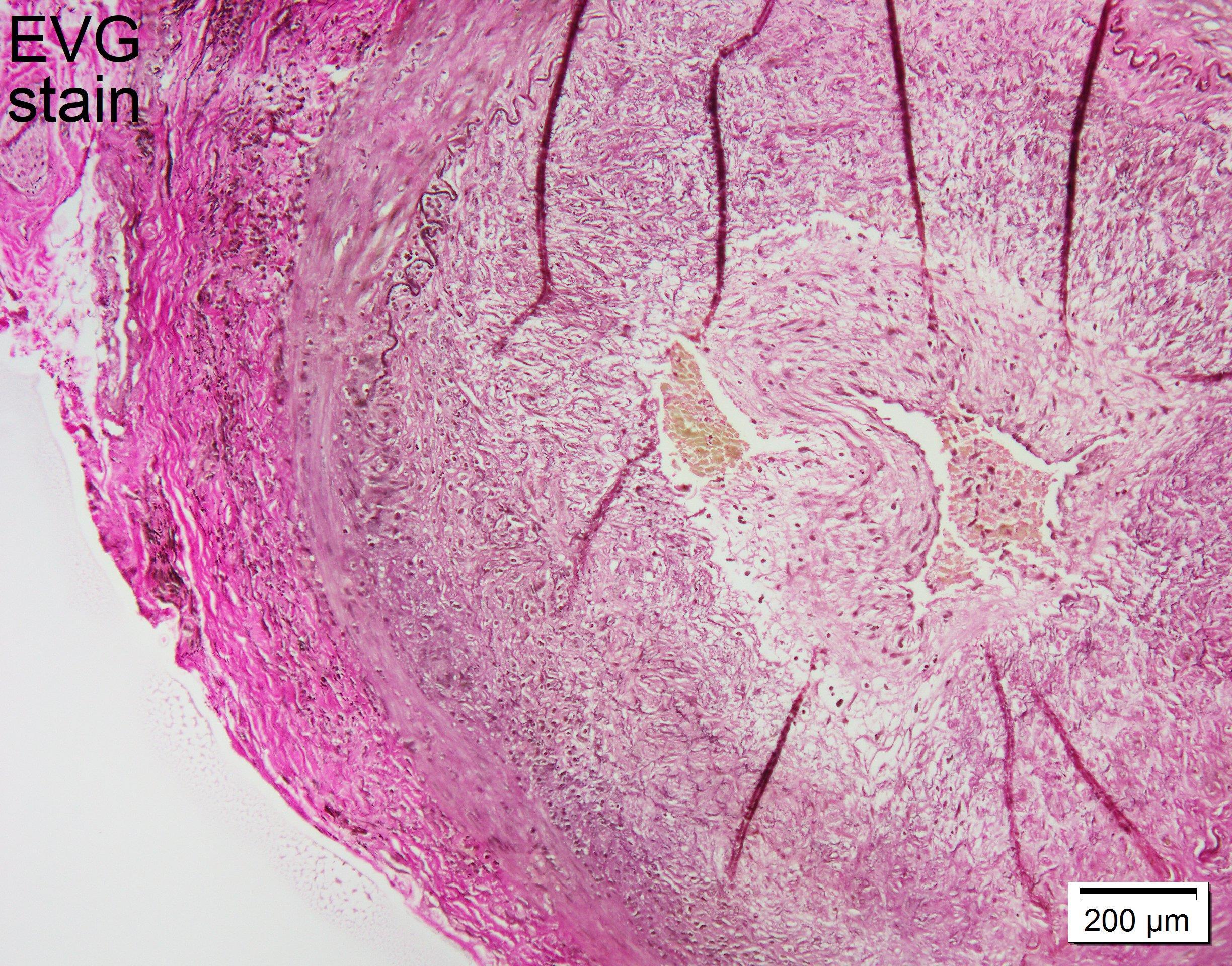
Figure 4: Patchy lymphoid infiltrates in adventitia with relatively preserved internal elastic membrane. (Click to enlarge.)
Our case involved an 81-year-old man who presented with symptoms typical for GCA but complicated by scalp necrosis. Scalp necrosis is a rare cutaneous manifestation of GCA, with only around 100 cases reported to date.4 It is usually a consequence of severe ischemia in the setting of delayed diagnosis and treatment of GCA.5 The presence of scalp necrosis in patients with GCA is associated with a higher frequency of vision loss and increased mortality.6 Early recognition and treatment with high-dose glucocorticoids are imperative.
Conclusion
It is important to recognize scalp necrosis in patients presenting with signs and symptoms of GCA so treatment can be initiated as soon as possible to prevent further complication and vision loss.
 Jude Al Qaqaa, MD, is an internal medicine resident at Lehigh Valley Health Network, Allentown, Pa., who will be applying for a rheumatology fellowship.
 Vladimir Falb, DO, is a rheumatology fellow at Lehigh Valley Health Network, Allentown, Pa. He completed his internal medicine residency at SUNY Downstate, Brooklyn, N.Y.
 Kaitlyn L Buzard, DO, is a rheumatologist at Lehigh Valley Health Network/Jefferson Health, Allentown, Pa. She has a special interest in vasculitis.
Acknowledgment
The authors thank Eugene Alexandrin, MD, for his assistance with the development of this article.
References
- Ameer MA, Sarosh V, Babak K. Giant Cell Arteritis (Temporal Arteritis) [updated 2024 May 2]. StatPearls [Internet]. Treasure Island, Fla.: StatPearls Publishing. 2025 Jan. https://tinyurl.com/22adszfd.
- Farina N, Tomelleri A, Campochiaro C, Dagna L. Giant cell arteritis: Update on clinical manifestations, diagnosis, and management. Eur J Intern Med. 2023 Jan;107:17–26.
- Ponte C, Martins-Martinho J, Luqmani RA. Diagnosis of giant cell arteritis. Rheumatology (Oxford). 2020 May 1;59 (Suppl 3):iii5–iii16.
- Idoudi S, Kahla MB, Mselmi F, et al. Scalp necrosis revealing severe giant-cell arteritis. Case Rep Med. 2020 Aug 14;2020:8130404.
- Prieto-Peña D, Castañeda S, Atienza-Mateo B, et al. A review of the dermatological complications of giant cell arteritis. Clin Cosmet Investig Dermatol. 2021 Mar 25;14:303–312.
- Tsianakas A, Ehrchen JM, Presser D et al. Scalp necrosis in giant cell arteritis: Case report and review of the relevance of this cutaneous sign of large-vessel vasculitis. J Am Acad Dermatol. 2009 Oct;61(4):701–706.