Video: Every Case Tells a Story| Webinar: ACR/CHEST ILD Guidelines in Practice

An official publication of the ACR and the ARP serving rheumatologists and rheumatology professionals

  • Conditions
    • Axial Spondyloarthritis
    • Gout and Crystalline Arthritis
    • Myositis
    • Osteoarthritis and Bone Disorders
    • Pain Syndromes
    • Pediatric Conditions
    • Psoriatic Arthritis
    • Rheumatoid Arthritis
    • Sjögren’s Disease
    • Systemic Lupus Erythematosus
    • Systemic Sclerosis
    • Vasculitis
    • Other Rheumatic Conditions
  • FocusRheum
    • ANCA-Associated Vasculitis
    • Axial Spondyloarthritis
    • Gout
    • Psoriatic Arthritis
    • Rheumatoid Arthritis
    • Systemic Lupus Erythematosus
  • Guidance
    • Clinical Criteria/Guidelines
    • Ethics
    • Legal Updates
    • Legislation & Advocacy
    • Meeting Reports
      • ACR Convergence
      • Other ACR meetings
      • EULAR/Other
    • Research Rheum
  • Drug Updates
    • Analgesics
    • Biologics/DMARDs
  • Practice Support
    • Billing/Coding
    • EMRs
    • Facility
    • Insurance
    • QA/QI
    • Technology
    • Workforce
  • Opinion
    • Patient Perspective
    • Profiles
    • Rheuminations
      • Video
    • Speak Out Rheum
  • Career
    • ACR ExamRheum
    • Awards
    • Career Development
  • ACR
    • ACR Home
    • ACR Convergence
    • ACR Guidelines
    • Journals
      • ACR Open Rheumatology
      • Arthritis & Rheumatology
      • Arthritis Care & Research
    • From the College
    • Events/CME
    • President’s Perspective
  • Search

Case Report: Lymphocytic Vasculitis of the Central Nervous System

Gbemisola Olayemi, MD, Evangeline Scopelitis, MD, & Jerald M. Zakem, MD  |  Issue: January 2019  |  January 17, 2019

Figure 8: Immunostaining for CD3 highlights the inflammatory T cell population permeating the vascular wall. CD20 was mostly negative.

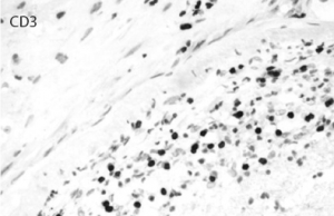
Courtesy of Maria-Beatriz Lopes, MD, University of Virginia Pathology

Discussion

PACNS is a poorly understood entity, and significant challenges remain for diagnosis and treatment. The pathogenesis is not well understood, and to date no evidence has been found to suggest a genetic predisposition. Various viral infections have been proposed as factors, such as varicella zoster virus, West Nile virus, Mycoplasma gallisepticum and HIV.6-14

ad goes here:advert-1
ADVERTISEMENT
SCROLL TO CONTINUE

In 1988, Calebrese and Mallek proposed diagnostic criteria for PACNS combining clinical, imaging and histology, which includes 1) acquired neurological deficit unexplained after complete evaluation, 2) diagnostic cerebral angiogram with narrowing of vessels suggestive of vasculitis, and 3) no evidence of systemic vasculitis or any other condition that could mimic the angiogram findings.15

These criteria were revised in 2009 by Birnbaum and Hellmann to either a definite or probable diagnosis of PACNS, with the main difference being confirmation of vasculitis on biopsy specimen to prevent patients with reversible vasoconstriction syndromes (RVCS) from being treated with cytotoxic therapy.16

ad goes here:advert-2
ADVERTISEMENT
SCROLL TO CONTINUE

Our patient fulfilled all three criteria for diagnosis of PACNS and had lymphocytic perivascular infiltrates on brain biopsy.

The characteristic histopathologic findings consist of inflammatory infiltration of vessel walls by T lymphocytes and activated macrophages, which undergo granulomatous differentiation with giant cell formation.

Different clinical subsets of PACNS include granulomatous angiitis of the central nervous system (GACNS), a rare subset of PACNS characterized by granulomatous angiitis confined to the brain. Patients present clinically with chronic insidious headaches, along with diffuse and focal neurologic deficits, often bilateral infarcts, as well as high-intensity T2-weighted fluid attenuation inversion recovery (FLAIR) lesions on MRI in the subcortical white matter and deep gray matter.

Atypical CNS vasculitis, another subset of PACNS, comprises multiple manifestations of PACNS that are clinically, radiologically or pathologically distinct from GACNS. The most frequent and heterogeneous subset of PACNS included in this subset are patients with specific presentations, such as lesions, or patients with pathologic findings of lymphocytic infiltration or necrotizing vasculitis with transmural fibrinoid necrosis rather than granulomatous angiitis.17

Benign angiopathy of the central nervous system (BACNS), which is now referred to as reversible cerebral vasoconstriction syndrome, was considered to be the third mimic of PACNS although not a true vasculitis.2

The median age of onset is 50 years, but PACNS may affect patients of all ages. The neurological manifestations are diverse, ranging from hyperacute to chronic and insidious. In a study of 101 patients, Salvarani et al. found patients who presented with subacute manifestations of diffuse CNS dysfunction and acute presentation were highly unusual.18

The most common initial symptoms are headache (63%) and cognitive impairment (50%). Focal symptoms usually appear later in the course of the disease and include hemiparesis (44%), stroke (40%), aphasia (28%), transient ischemic attack (28%), ataxia (19%), seizures (16%), dysarthria (15%) and blurred vision or decreased visual acuity (11%). Infrequent manifestations, occurring in less than 10% of patients, include intracranial hemorrhage; amnesic syndrome; and spinal cord manifestations, such as paraparesis, quadriparesis, Parkinsonism, vertigo, dizziness or cranial nerve palsy; and most patients have multiple manifestations.

No definite laboratory abnormality is diagnostic. The diagnosis of PACNS is challenging because of the nonspecific clinical presentation, the lack of highly specific laboratory and radiologic tests, and the difficulty of obtaining pathologic material. In the majority of published case series of PACNS, abnormal cerebrospinal fluid (CSF) findings have been reported; both CSF pleocytosis and protein elevation are frequent.19 Obtaining appropriate CSF cultures, microbiologic stains, cytology and flow cytometry is crucial to rule out infectious and neoplastic disease.

MRI is the neuroimaging modality of choice for patients with suspected PACNS, and MRI findings are abnormal in 90–100% of patients. MRI may demonstrate ischemic and hemorrhagic lesions of different ages, leukencephalopathies, tumor-like lesions or gadolinium enhancement of the meninges.20 Infarcts may be seen in approximately 50% of cases; when present, infarcts are usually seen bilaterally in multiple-vessel tributaries affecting the cortex, as well as the subcortex. Gadolinium enhancement may occur in as many as one-third of cases; leptomeningeal enhancement may occur in 10–15% of cases, and when present in the nondominant lobe, may represent an ideal site for biopsy.21

Angiography sometimes demonstrates bilateral vessel stenoses or occlusions consistent with an angiitis. In some cases, patients with histologically proved PACNS have an entirely normal angiogram. The angiographic pattern considered diagnostic of vasculitis is often caused by reversible vasoconstriction syndromes associated with drugs, migraine, hypertension, eclampsia or the postpartum period.19,22

Patients must be evaluated for systemic vasculitis, which is characterized by the presence of constitutional symptoms and serological markers indicating systemic inflammation. The inflammatory process in PACNS is limited to the CNS.

Several diseases can mimic PACNS, including reversible cerebral vasoconstriction syndrome, premature intracranial atherosclerosis, fibromuscular dysplasia, secondary cerebral vasculitis, malignancy, infections (e.g., VZV, HIV, hepatitis C, tuberculosis), other multi-system inflammatory disorders, such as sarcoidosis, moya­moya disease, genetic conditions (e.g., CADASIL), posterior reversible encephalopathy syndrome (PRES), chronic hypertension (microvascular cerebral ischemia), demyelinating diseases and others with multifocal cerebral thromboembolism. Other rare syndromes resembling cerebral vasculitis include reversible posterior leukoencephalopathy syndrome, MELAS (mitochondrial encephalomyopathy, lactic acidosis and stroke-like episodes), ma­lignant intravascular lymphomatosis, Degos disease, amyloid angiopathy, Fabry’s disease, pseudoxanthoma elasticum, lipohyalinosis and storage diseases.2,20

Brain biopsy is the gold standard for diagnosis to confirm and exclude other etiologies. In a study done by Alrawi et al., out of 61 patients referred for biopsy to test for suspected PACNS, alternative diagnoses were established in 24 cases (39%); 12 patients had infections, including three patients with brain abscesses, and eight patients had malignant neoplasms, including six with primary CNS lymphoma.23 CNS vasculitis is a patchy disease that affects vessels in a skipped and segmental pattern, which limits the sensitivity of the procedure, and as many as 25% of the biopsies were falsely negative.23 To improve the diagnostic yield, targeted biopsies should be performed with inclusion of leptomeninges.17,24

Unfortunately, no randomized studies of PACNS have been done, and thus, all information on treatment is based on retrospective observational data and clinical experience.

Treatment recommendations are derived from the protocols for systemic vasculitides with severe organ involvement in which a combination of steroids with cyclophosphamide is recommended.20 The majority of case series suggest a high degree of good outcomes when patients are treated with glucocorticoids or glucocorticoids and cyclophosphamide.

After remission is achieved, maintenance therapy with azathioprine or mycophenolate mofetil—and, rarely, methotrexate, given its low CNS penetration—is initiated.2 Some reports indicate that combining mycophenolate mofetil and steroids allows disease control with disappearance of the neurological abnormalities, restoration of normal daily activities and dramatic improvement in brain MRI abnormalities.25 Some case reports used rituximab as the initial treatment for CNS vasculitis, and TNF alpha inhibitors are being used for treatment of CNS vasculitis resistant to immunosuppressive treatment.18,26,27

Page: 1 2 3 4 5 6 7 | Single Page
Share: 

Filed under:ConditionsVasculitis Tagged with:central nervous system vasculitiscyclophosphamideMethylprednisolonePrimary Angiitis of the Central Nervous System

Related Articles

    Meet the Challenge of Primary CNS Vasculitis

    September 1, 2011

    Diagnosis and treatment of this rare and poorly understood condition

    Rheumatology Case Report: When Moyamoya Disease Mimicks Primary Central Nervous System Vasculitis

    November 16, 2015

    Case report: A 60-year-old Hispanic male with poorly controlled hypertension was sent from the primary care clinic for evaluation of malignant hypertension with a systolic blood pressure above 200 mmHg. His symptoms at the time of presentation included episodic confusion, worsening vision and an unsteady gait. A head computed tomography (CT) scan showed a subacute…

    7 Key Insights Into the Evaluation of Central Nervous System Vasculitis

    May 13, 2021

    Many a rheumatology consult has centered on a perplexing question: Does this patient have central nervous system (CNS) vasculitis? At the 2021 ACR State-of-the-Art Clinical Symposium, Rula Hajj-Ali, MD, FACP, professor of medicine and associate director of vasculitis care and research, Cleveland Clinic Lerner College of Medicine, discussed this topic in detail, providing a series…

    2014 ACR/ARHP Annual Meeting: Diagnosing PACNS and Its Mimics

    February 1, 2015

    Accurate diagnosis for primary angiitis of the central nervous system requires close scrutiny, team effort

  • About Us
  • Meet the Editors
  • Issue Archives
  • Contribute
  • Advertise
  • Contact Us
  • Copyright © 2025 by John Wiley & Sons, Inc. All rights reserved, including rights for text and data mining and training of artificial technologies or similar technologies. ISSN 1931-3268 (print). ISSN 1931-3209 (online).
  • DEI Statement
  • Privacy Policy
  • Terms of Use
  • Cookie Preferences