Video: Every Case Tells a Story| Webinar: ACR/CHEST ILD Guidelines in Practice

An official publication of the ACR and the ARP serving rheumatologists and rheumatology professionals

  • Conditions
    • Axial Spondyloarthritis
    • Gout and Crystalline Arthritis
    • Myositis
    • Osteoarthritis and Bone Disorders
    • Pain Syndromes
    • Pediatric Conditions
    • Psoriatic Arthritis
    • Rheumatoid Arthritis
    • Sjögren’s Disease
    • Systemic Lupus Erythematosus
    • Systemic Sclerosis
    • Vasculitis
    • Other Rheumatic Conditions
  • FocusRheum
    • ANCA-Associated Vasculitis
    • Axial Spondyloarthritis
    • Gout
    • Psoriatic Arthritis
    • Rheumatoid Arthritis
    • Systemic Lupus Erythematosus
  • Guidance
    • Clinical Criteria/Guidelines
    • Ethics
    • Legal Updates
    • Legislation & Advocacy
    • Meeting Reports
      • ACR Convergence
      • Other ACR meetings
      • EULAR/Other
    • Research Rheum
  • Drug Updates
    • Analgesics
    • Biologics/DMARDs
  • Practice Support
    • Billing/Coding
    • EMRs
    • Facility
    • Insurance
    • QA/QI
    • Technology
    • Workforce
  • Opinion
    • Patient Perspective
    • Profiles
    • Rheuminations
      • Video
    • Speak Out Rheum
  • Career
    • ACR ExamRheum
    • Awards
    • Career Development
  • ACR
    • ACR Home
    • ACR Convergence
    • ACR Guidelines
    • Journals
      • ACR Open Rheumatology
      • Arthritis & Rheumatology
      • Arthritis Care & Research
    • From the College
    • Events/CME
    • President’s Perspective
  • Search

Case Report: Perplexing Pulmonary Nodules

Monsoon Rashid, DO, & Deana Lazaro, MD  |  Issue: March 2022  |  March 14, 2022

Figure 1: Computed Tomography

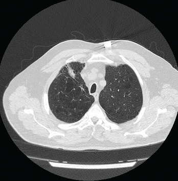
Figure 1: Computed Tomography VGstockstudio / shutterstock.com

Pulmonary nodules are com­mon; most are benign, but the differential diagnosis is broad and includes life-threatening possibilities.1 Our patient is a former smoker who has a history of a complex autoimmune disease and multiple pulmonary nodules. This case was challenging, but clinical, radiographic and histologic clues helped lead to the correct diagnosis.

Case Presentation

The patient is a 74-year-old man with a past medical history remarkable for hypertension, hyperlipidemia, gastroesophageal reflux disease, emphysema, asbestos exposure and seropositive (rheumatoid factor and anti-cyclic citrullinated protein antibody) rheumatoid arthritis/scleroderma/systemic lupus erythematosus (SLE) overlap syndrome. He has a 100 pack-year smoking history, although he quit smoking 22 years ago.

ad goes here:advert-1
ADVERTISEMENT
SCROLL TO CONTINUE

The patient was diagnosed with sero­positive rheumatoid arthritis 15 years ago. His initial treatment was 15 mg of oral methotrexate weekly and 40 mg of subcutaneous adalimumab every 14 days. His methotrexate was tapered to 10 mg weekly, and the patient did well.

His disease course was complicated by the development of bulky rheumatoid nodules on the extensor surfaces of his elbows. After surgical removal of the nodules, he was taken off methotrexate and switched to 1 g of sulfasalazine twice daily to reduce the risk of rheumatoid nodulosis. Due to his moderately active disease, his therapy was changed from sulfasalazine to 20 mg of leflunomide daily.

ad goes here:advert-2
ADVERTISEMENT
SCROLL TO CONTINUE

The patient stopped taking adalimumab because he felt well.

The pulmonary nodules in rheumatoid pneumoconiosis develop cavitations or calcifications in 10% of cases.

Four years ago, the patient complained of Raynaud’s phenomenon. A physical examination revealed skin tightness from his fingertips to his proximal interphalangeal joints.

Serologic tests were positive for anti-nuclear antibody (ANA; 1:160, in a speckled pattern) and anti-centromere antibody (ACA; 2.8 AI; reference range [RR]: 0.0–0.9 AI). Tests for anti-SSA/Ro antibody, anti-SSB/La antibody, anti-nuclear ribonucleoprotein (anti-RNP) antibody, anti-Smith antibody and anti-topoisomerase I antibody were negative.

Two years ago, the patient had a flare of inflammatory arthritis, and treatment was started with 125 mg of subcutaneous abatacept weekly.

A few months later, the patient developed dyspnea due to serositis. He had no fever, cough, weight loss, night sweats or joint pain. A QuantiFERON gold test was negative for tuberculosis.

Results of pleural fluid studies included: albumin of 3.1 g/dL (RR: 3.5–5.0 g/dL), white blood cell count of 2535/µL (RR: 4,000–11,000/µL) with 44% neutrophils, glucose of 2 mg/dL (RR: 60–99 mg/dL) and a lactate dehydrogenase of 2,308 U/L (RR: 140–280 U/L).

Cultures for bacteria, fungi and acid-fast bacilli showed no growth. Cytology of the fluid revealed no malignant cells. The pleural fluid studies were consistent with an exudative, rheumatoid pleural effusion.

The patient experienced atypical chest pain. Evaluation for acute coronary syndrome was negative.

One month later, chest imaging showed a pericardial effusion and a repeat echo­cardiogram was consistent with tamponade. A pericardial window was performed to manage cardiac tamponade, and biopsy of the pericardium revealed fibrous tissue with congestion, chronic inflammation and granulation tissue.

Cultures of the pericardial fluid for bacteria, acid-fast bacilli and fungi were unrevealing. Anti-proteinase-3 (PR3) antibody was 4.3 U/mL (RR: 0–3.5 U/mL). The patient also had reduced C4 complement, 6.9 mg/dL (RR: 9–36 mg/dL), and IgM anti-cardiolipin antibody was mildly elevated at 39 U/mL (RR: 1–12 U/mL). The patient had no joint inflammation or renal disease at that time.

The patient had a history of urticaria after taking non-steroidal anti-inflammatory drugs; therefore, he was treated with 10 mg of prednisone daily and 0.6 mg of colchicine twice daily. Sulfasalazine was discontinued because serositis was reported as a possible side effect of this drug in case reports, and 200 mg of hydroxychloroquine twice daily was initiated.

The following tests were performed: anti-double-stranded DNA (anti-dsDNA) antibody <1 IU/mL (RR: 0–9 IU/mL), anti-RNP antibodies <0.2 AI (RR: 0.0–0.9 AI), anti-nuclear cytoplasmic antibodies <1:20 (RR: <1:20 titer), anti-myeloperoxidase antibody <9.0 U/mL (RR: 0.0–9.0 U/mL) and anti-proteinase-3 antibody <3.5 U/mL (RR: 0.0–3.5 U/mL).

Pulmonary function studies were consistent with moderate obstructive lung disease and moderate decrease in diffusion capacity. Computed tomography (CT) scan of the thorax revealed severe emphysematous changes, a 1.8 cm x 1.6 cm pleural-based nodule in the anterior aspect of the right upper lobe and multiple sub-centimeter pulmonary nodules (see Figure 1).

Two percutaneous transthoracic biopsies of the largest left upper lobe and right upper lobe nodules showed patchy inter­stitial fibrosis and anthracotic pigmented macrophages. No malignancy or granuloma was identified on histology, and cultures for bacteria were negative.

Page: 1 2 3 | Single Page
Share: 

Filed under:Conditions Tagged with:Caplan Syndromecase reportrheumatoid pneumoconiosis

Related Articles

    Multidisciplinary Guidance for Pulmonary Disease in Sjögren’s Syndrome

    February 2, 2022

    Lung disease is common, variable and frequently underdiagnosed in patients with Sjögren’s syndrome. An expert pulmonologist and a rheumatologist discussed the diagnosis and management of pulmonary manifestations of Sjögren’s syndrome during ACR Convergence 2021.

    Labored Breathing

    January 1, 2007

    Strategies to diagnose and manage dyspnea in the scleroderma patient

    Case Report: Interstitial Lung Disease with Positive ANCA Test

    October 13, 2015

    “Worst of all is the pain in my calves,” she said. “It feels like burning deep inside.” So began my first encounter with a 69-year-old woman who was referred to rheumatology clinic for evaluation of two months of constitutional symptoms and a positive ANCA test, which had been ordered by her primary care doctor. Her…

    Case Study: 66-Year-Old Man with Visual Loss, Headache, Hematuria, Chronic Sinusitis

    May 8, 2012

    A 66-year-old man presented to the hospital with left-sided visual loss, headache, hematuria, and symptoms of a chronic sinusitis.

  • About Us
  • Meet the Editors
  • Issue Archives
  • Contribute
  • Advertise
  • Contact Us
  • Copyright © 2025 by John Wiley & Sons, Inc. All rights reserved, including rights for text and data mining and training of artificial technologies or similar technologies. ISSN 1931-3268 (print). ISSN 1931-3209 (online).
  • DEI Statement
  • Privacy Policy
  • Terms of Use
  • Cookie Preferences