Video: Every Case Tells a Story| Webinar: ACR/CHEST ILD Guidelines in Practice

An official publication of the ACR and the ARP serving rheumatologists and rheumatology professionals

  • Conditions
    • Axial Spondyloarthritis
    • Gout and Crystalline Arthritis
    • Myositis
    • Osteoarthritis and Bone Disorders
    • Pain Syndromes
    • Pediatric Conditions
    • Psoriatic Arthritis
    • Rheumatoid Arthritis
    • Sjögren’s Disease
    • Systemic Lupus Erythematosus
    • Systemic Sclerosis
    • Vasculitis
    • Other Rheumatic Conditions
  • FocusRheum
    • ANCA-Associated Vasculitis
    • Axial Spondyloarthritis
    • Gout
    • Psoriatic Arthritis
    • Rheumatoid Arthritis
    • Systemic Lupus Erythematosus
  • Guidance
    • Clinical Criteria/Guidelines
    • Ethics
    • Legal Updates
    • Legislation & Advocacy
    • Meeting Reports
      • ACR Convergence
      • Other ACR meetings
      • EULAR/Other
    • Research Rheum
  • Drug Updates
    • Analgesics
    • Biologics/DMARDs
  • Practice Support
    • Billing/Coding
    • EMRs
    • Facility
    • Insurance
    • QA/QI
    • Technology
    • Workforce
  • Opinion
    • Patient Perspective
    • Profiles
    • Rheuminations
      • Video
    • Speak Out Rheum
  • Career
    • ACR ExamRheum
    • Awards
    • Career Development
  • ACR
    • ACR Home
    • ACR Convergence
    • ACR Guidelines
    • Journals
      • ACR Open Rheumatology
      • Arthritis & Rheumatology
      • Arthritis Care & Research
    • From the College
    • Events/CME
    • President’s Perspective
  • Search

Case Report: Possible Overlapping Vasculitis & Ulcerative Colitis

Julia Jing-ou Tan, MD, Mohammad Bardi, MD, & Natasha Dehghan, MD  |  Issue: January 2020  |  January 17, 2020

The patient had nodular pus-filled cystic lesions with violaceous borders on his left ear, right cheek, fingers and legs.

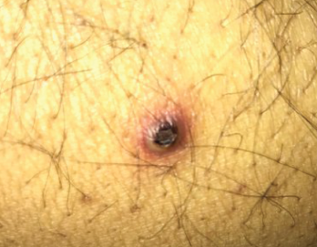
Figure 1. The patient had nodular pus-filled cystic lesions with violaceous borders on his left ear, right cheek, fingers and legs.

A 42-year-old man with a history of ulcerative colitis (UC), primary sclerosing cholangitis (PSC) and chronic sinusitis was referred to a rheumatologist to evaluate for a possible diagnosis of systemic vasculitis. This patient had developed new skin lesions, gingival hypertrophy and ulcerating tracheobronchitis, concerning for possible granulomatosis with polyangiitis (GPA).

Since 1994, the patient had sinusitis with occasional bloody discharge and nasal turbinate hypertrophy demonstrated on imaging. He was followed by an otolaryngologist, and his symptoms were well controlled with budesonide nasal rinses. In 1996, the patient presented to the hospital with abdominal pain and bloody diarrhea. A subsequent colonoscopy and biopsy confirmed a diagnosis of UC. He was treated with mesalazine, and in 1998, when he developed PSC, ursodiol was added.

ad goes here:advert-1
ADVERTISEMENT
SCROLL TO CONTINUE

Overall, his UC was well controlled until 2012. At that time, he began requiring intermittent steroids for UC flares, and in 2015, his gastroenterologist started treatment with vedolizumab.

In late 2016, the patient developed nodular, pus-filled cystic lesions with violaceous borders on his left ear, right cheek, fingers and legs (see Figure 1). Biopsy of these cutaneous manifestations were suggestive of neutrophilic folliculitis, a variant of pyoderma gangrenosum.1

ad goes here:advert-2
ADVERTISEMENT
SCROLL TO CONTINUE

Around the same time, the patient also started having severely painful, bleeding gingival hypertrophy (see Figure 2). Based on the appearance of his gums, this symptom was thought to be strawberry gingivitis, a rare, but classic, feature of GPA.2 Consequently, the possibility of an overlapping vasculitis concurrently occurring with the patient’s inflammatory bowel disease (GPA-IBD overlap) was suggested. As a result, azathioprine and prednisone were added to the patient’s medication regimen.

Figure 2. The patient presented with severe, painful, bleeding gingival hypertrophy.

Figure 2. The patient presented with severe, painful, bleeding gingival hypertrophy.

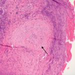
After review with multiple specialists, gingival biopsies in January 2017 demonstrated pyostomatitis vegetans, which is a known extra-intestinal manifestation of IBD.

Sinus biopsies were also performed and did not show any evidence of vasculitis. Further, serologic evaluation during this period of diagnostic inquiry was variable. In March 2016, the patient was positive for C-ANCA (anti-neutrophilic cytoplasmic antibodies), but was PR3-ANCA and MPO-ANCA negative. In February 2017, he had an atypical P-ANCA (perinuclear ANCA) with a PR3-ANCA of 49.6 (normal <20). In March 2017, all ANCA serologies were negative.

Over the next eight months, the patient was treated with increasing doses of azathio­prine and prednisone, up to 150 mg once daily and 50 mg once daily, respectively. But his pyostomatis vegetans failed to respond, and he continued to have significantly distressing gum pain and bleeding.

In September 2017, in an attempt to control this symptom, he was switched from vedolizumab to infliximab. This change initially improved his gingival manifestations after the first dose; however, subsequent doses, including dose escalations, did not achieve the same therapeutic effect. Additionally, his previously well-controlled gastrointestinal symptoms flared, and the decision was made to switch him back to vedolizumab.

In March 2018, the patient was admitted to the hospital with decompensated liver cirrhosis and an acute portal vein thrombosis. He underwent liver transplant and was discharged home.

Page: 1 2 3 4 | Single Page
Share: 

Filed under:ConditionsVasculitis Tagged with:case reportulcerative colitis

Related Articles
    Figure 2: Renal Biopsy

    The Classification & Diagnosis of Granulomatosis with Polyangiitis

    August 16, 2018

    Based on the classification system developed by the Chapel Hill Consensus Conference, anti-neutrophil cytoplasmic antibody (ANCA) associated vasculitis is defined as a necrotizing vasculitis involving small vessels that is associated with myeloperoxidase (MPO) ANCA or proteinase 3 (PR3) ANCA and displays minimal immune deposits. The mechanism behind the pathogenesis of ANCA-associated vasculitis is not fully…

    Figure 1. A nasal biopsy shows intimal infiltration of the small blood vessels (black arrow).

    Case Illustrates the Difficulty Diagnosing Granulomatosis with Polyangiitis

    June 21, 2018

    Granulomatosis with polyangiitis (GPA) was first described in the British Medical Journal in 1897 by Scottish otolaryngologist Peter McBride.1 GPA is a relatively rare, systemic necrotizing vasculitis that can make diagnosis challenging. The incidence has been estimated anywhere between two and 12 cases per million.2 GPA mainly affects adults between the ages of 45 and…

    Case Report: Hydralazine-Induced ANCA-Associated Vasculitis

    February 16, 2021

    Hydralazine has been in use as a treatment for hypertension, most notably in heart failure patients, since 1951.1 The drug is a known cause of autoimmune disease, most specifically hydralazine-induced lupus.  Hydralazine-induced lupus occurs in 7–13% of those taking the medication.2-4 It often presents with constitutional symptoms, arthritis/arthralgias, cutaneous lesions, sero­sitis, myalgias and/or hepatomegaly. Features…

    Gut Feeling: A Comprehensive Look at the Pathogenesis, Management & Treatment of Inflammatory Bowel Disease

    May 24, 2021

    Patients with autoimmune diseases, such as spondyloarthritis, are at risk of developing inflammatory bowel disease. Here are considerations for its management and treatment.

  • About Us
  • Meet the Editors
  • Issue Archives
  • Contribute
  • Advertise
  • Contact Us
  • Copyright © 2025 by John Wiley & Sons, Inc. All rights reserved, including rights for text and data mining and training of artificial technologies or similar technologies. ISSN 1931-3268 (print). ISSN 1931-3209 (online).
  • DEI Statement
  • Privacy Policy
  • Terms of Use
  • Cookie Preferences