Video: Superheroes, Secret Identities & You| Webinar: ACR/CHEST ILD Guidelines in Practice

An official publication of the ACR and the ARP serving rheumatologists and rheumatology professionals

  • Conditions
    • Axial Spondyloarthritis
    • Gout and Crystalline Arthritis
    • Myositis
    • Osteoarthritis and Bone Disorders
    • Pain Syndromes
    • Pediatric Conditions
    • Psoriatic Arthritis
    • Rheumatoid Arthritis
    • Sjögren’s Disease
    • Systemic Lupus Erythematosus
    • Systemic Sclerosis
    • Vasculitis
    • Other Rheumatic Conditions
  • FocusRheum
    • ANCA-Associated Vasculitis
    • Axial Spondyloarthritis
    • Gout
    • Lupus Nephritis
    • Psoriatic Arthritis
    • Rheumatoid Arthritis
    • Systemic Lupus Erythematosus
  • Guidance
    • Clinical Criteria/Guidelines
    • Ethics
    • Legal Updates
    • Legislation & Advocacy
    • Meeting Reports
      • ACR Convergence
      • Other ACR meetings
      • EULAR/Other
    • Research Rheum
  • Drug Updates
    • Analgesics
    • Biologics/DMARDs
  • Practice Support
    • Billing/Coding
    • EMRs
    • Facility
    • Insurance
    • Technology
      • Information Technology
      • Apps
    • QA/QI
    • Workforce
  • Opinion
    • Patient Perspective
    • Profiles
    • Rheuminations
      • Video
    • Speak Out Rheum
  • Career
    • ACR ExamRheum
    • Awards
    • Career Development
      • Education & Training
    • Certification
  • ACR
    • ACR Home
    • ACR Convergence
    • ACR Guidelines
    • Journals
      • ACR Open Rheumatology
      • Arthritis & Rheumatology
      • Arthritis Care & Research
    • From the College
    • Events/CME
    • President’s Perspective
  • Search

Case Report: Pulmonary Artery Stenosis Secondary to Granulomatosis with Polyangiitis

Sambhawana Bhandari, MD, & Colin Diffie, MD  |  Issue: August 2025  |  August 7, 2025

The 2012 revised International Chapel Hill Consensus Conference classified vasculitis on the basis of the size of predominantly affected vessels, as large-, medium- or small-vessel vasculitis.1 Anti-neutrophil cytoplasmic antibody (ANCA) associated vasculitis is a systemic autoimmune disease that primarily involves necrotizing inflammation of small- and medium-sized vessels. However, ANCA-associated vasculitis can occasionally extend to large vessels, including the aorta, supra-aortic trunks and pulmonary arteries, challenging its classical categorization.2,3 The main subtypes of ANCA-associated vasculitis are granulomatosis with polyangiitis (GPA), microscopic polyangiitis (MPA) and eosinophilic granulomatosis with polyangiitis (EGPA).

Pulmonary artery stenosis, although rare, is an important clinical entity that can be either congenital or acquired. Acquired causes include vasculitis, pulmonary thromboembolism and extrinsic compression by mediastinal masses or lymphadenopathy.4,5 Large-vessel vasculitides, such as Takayasu arteritis, giant cell arteritis and Behçet’s disease, are well established causes of pulmonary artery involvement. In contrast, large-vessel involvement in ANCA-associated vasculitis, including pul­monary artery stenosis, is far less common and often under-recognized.5

ad goes here:advert-1
ADVERTISEMENT
SCROLL TO CONTINUE

Here, we report a rare case of bilateral pulmonary artery stenosis secondary to GPA, underscoring the importance of recognizing large-vessel involvement as a potential manifestation of ANCA-associated vasculitis.

Case Presentation

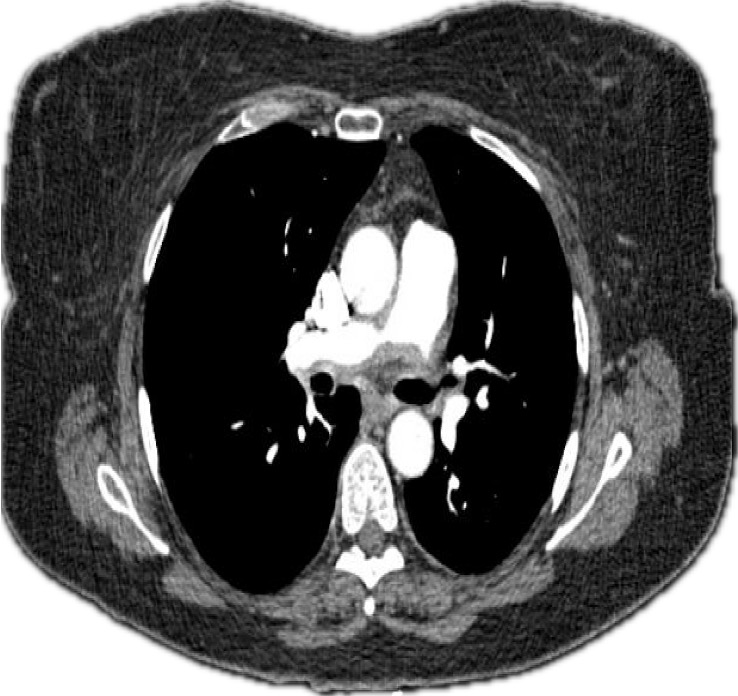
Figure 1: CTA of the chest showing stenosis of the right pulmonary artery. (Click to enlarge.)

A 62-year-old woman with a history of well-controlled type 2 diabetes mellitus presented with a progressive constellation of symptoms that began in early 2021. A nurse herself, initially she reported “hearing a bruit” in her chest without associated symptoms.

ad goes here:advert-2
ADVERTISEMENT
SCROLL TO CONTINUE

Around this time, she developed recurrent nasal congestion with bloody discharge and sinus infections. Over the ensuing months, she experienced progressive left-side hearing loss, recurrent otitis media requiring tympanoplasty and arthralgias. A few months later, she noted progressive bilateral leg swelling. Her physical therapist identified worsening exertional dyspnea and auscultated a heart murmur. She also experienced worsening fatigue and noted scattered petechiae on her legs, arms, and abdomen.

Given her worsening dyspnea, leg swelling and palpitations, she was referred to a cardiologist. A trans­thoracic echocardiogram revealed severe pulmonary hypertension with an estimated right ventricular systolic pressure (RVSP) of 102 mmHg, right ventricular dilation and a normal ejection fraction.

Holter monitoring ruled out arrhythmias. A computed tomography and angiogram (CTA) of the chest revealed ill-defined, soft-tissue thickening surrounding the right and left main pulmonary arteries, with moderate stenosis (see Figures 1 and 2).

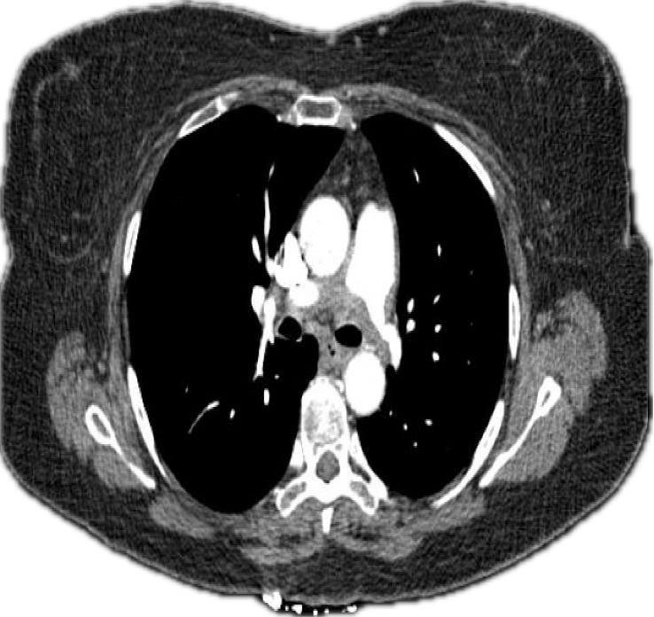
Figure 2: CTA of the chest showing stenosis of the left pulmonary artery.

Positron emission tomography-computed tomography (PET-CT) imaging demonstrated infiltrative tissue at the pulmonary artery outflow tract and increased metabolic activity in the proximal pulmonary arteries (see Figures 3 and 4).

Laboratory tests were notable for a positive ANCA at a titer of 1:640, P-ANCA pattern, and with a positive immunoassay for myeloperoxidase (MPO) >8. Anti-nuclear antibody (ANA), extractable nuclear antigen (ENA), double-stranded DNA (dsDNA) and eosinophil counts were negative or normal. Urinalysis revealed microscopic hematuria (2+ blood, >50 red blood cells per high-power field) with normal protein levels and protein-creatinine ratio. Other laboratory results, including white blood cell count, hemoglobin, liver function tests and creatinine, were unremarkable.

The patient was started on 60 mg of prednisone daily, 30 mg of avacopan twice daily and 1 g of rituximab every two weeks for two doses. She reported symptomatic improvement, including resolution of dyspnea, rash, sinus symptoms and constitutional complaints.

A ventilation-perfusion scan demonstrated diffuse and focal areas of decreased perfusion in the left lung. Right heart catheterization (RHC) confirmed proximal right pulmonary artery stenosis, with a peak gradient of 24 mmHg, and pulmonary artery systolic pressure (PASP) of 72 mmHg, with a mean pulmonary artery pressure (mPAP) of 32 mmHg. Left and right heart filling pressures and cardiac output were normal.

Figure 3 (transverse view): PET-CT demonstrating increased metabolic activity in the proximal pulmonary arteries. (Click to enlarge.)

With treatment, the patient experienced symptomatic improvement as prednisone was tapered. A follow-up PET scan showed reduced inflammation in the pulmonary artery outflow tract and proximal pulmonary arteries. Repeat chest CT angiography was stable, and pulmonary function tests and CT imaging of the sinuses were normal.

Subsequently, she underwent bilateral pulmonary artery stenting, leading to a significant improvement in pulmonary artery systolic pressure (PASP) from 72 mmHg to 35 mmHg. She continued to do well clinically on follow-up.

Discussion

The involvement of large vessels, such as the pulmonary artery, in ANCA-associated vasculitis is a rare but clinically significant manifestation. Pulmonary artery involvement can lead to such complications as progressive stenosis, pulmonary hypertension and right-side heart failure.

A multi-center case-control study by Monghal et al. found the aorta was the most commonly affected vessel, with involvement in 69% of cases. Other affected vessels included the supra-aortic trunks (35%), lower-limb arteries (19%), mesenteric arteries (19%), renal arteries (15%) and upper-limb arteries (8%).3 Pulmonary artery involvement was not noted in this cohort. However, multiple case reports have documented pulmonary artery involvement in ANCA-associated vasculitis, indicating that although rare, it is a recognized clinical entity.

Figure 4 (coronal view): PET-CT demonstrating increased metabolic activity in the proximal pulmonary arteries. (Click to enlarge.)

We conducted a literature review using the following search termi­nology: (“Granulomatosis with Polyangiitis” [MeSH] OR “Microscopic Polyangiitis” [MeSH] OR “Anti-Neutrophil Cyto­plasmic Antibody-Associated Vasculitis” [MeSH] OR “ANCA-associated vasculitis” OR “Wegener’s granulo­matosis” or myelo­peroxidase antibody OR pro­teinase 3 antibody) AND (“Pulmonary Artery” [MeSH] OR “Pulmonary Artery Stenosis” [MeSH] OR “Pulmonary Arteritis” OR “pulmonary artery vasculitis”).

This search yielded 45 results, from which we reviewed eight relevant articles.2,4-10 Including our patient, we reviewed a total of nine patients.

Table 1 (see below) summarizes reported cases, highlighting the diverse presentations, diagnostic challenges and therapeutic approaches associated with pul­monary artery stenosis in ANCA-associated vasculitis.

The age of affected patients ranged from 22 to 62 years, with a majority being women (78%). Most patients had classical ENT (ear, nose and throat) symptoms prior to the diagnosis of pulmonary stenosis, with duration ranging from three to six years. In some cases, pulmonary artery stenosis was synchronous with the diagnosis of GPA; in one patient, it was discovered 21 years after the diagnosis of GPA.

Diagnosis relied on clinical features, serological tests, imaging and histopathology. C-ANCA with anti-proteinase 3 (PR3) antibody was detected in four of seven patients. P-ANCA with MPO antibody was detected in three of seven patients; in two cases, this information was unavailable. Imaging modalities, such as CT pulmonary angiogram, conventional angiogram, echocardiography and FDG-PET were pivotal. Angiograms were mainly helpful in diagnosing luminal abnormalities; FDG-PET and cardiac MRI were useful for detecting vessel wall abnormalities, and ECHO/RHC for evaluating pulmonary hypertension.

The pulmonary artery lesions typically presented as severe stenosis involving the proximal or main branches of the pulmonary arteries, although distal involvement was also noted in one patient.2 Among the nine cases reviewed, four confirmed the diagnosis with a histopathology showing pulmonary arteritis secondary to GPA.4,6,7,10

A case report by Doyle et al. noted that imaging of the pulmonary arteries with conventional imaging modalities, such as CTPA (computed tomography pulmonary angiography) or conventional pulmonary angiogram, is identical between arteritis and chronic thromboembolic disease and often needs a histopathological diagnosis for confirmation, which can unfortunately be challenging due to surgical complications and isn’t always obtained.7 Use of multimodal imaging techniques, including CTPA, conventional angiogram, echocardiogram and FDG-PET can be useful in diagnosing pulmonary stenosis and assessing the severity of pulmonary hypertension.

Pulmonary artery involvement can be an isolated finding with GPA, without involvement of any other large vessels. Two case reports noted involvement of the ascending aorta, and one case reported involvement of the internal thoracic arteries and coronary arteries, along with the ascending aorta and pulmonary artery.5,9

Although one might argue that our patient could have an overlap of GPA and Takayasu’s arteritis, she is in her 60s, which is not the typical demographic, and she didn’t have involvement of the aortic arch, subclavian or carotid vessels on imaging.

Immunosuppressive therapy, including glucocorticoids and such agents as cyclophosphamide or rituximab, forms the cornerstone of treatment for ANCA-associated vasculitis with large-vessel involvement. However, its efficacy in addressing pulmonary artery stenosis remains variable. In the reviewed cases, immunosuppressive therapy improved the severity of stenosis in two of five medically managed cases, with one case remaining stable. Despite optimal medical management, two cases required pulmonary artery stenting due to progressive/persistent stenosis. This variability is likely explained by the underlying pathology: While immunosuppression effectively reduces active inflammation (arteritis), stenosis frequently results from irreversible fibrosis and vascular remodeling, which may not respond to anti-inflammatory or immuno­suppressive agents.

ACR guidelines for Takayasu’s arteritis favor medical management over procedural management.11 However, in select cases, procedural interventions, such as pulmonary artery stenting, play a critical role in managing severe stenosis or refractory symptoms. Pulmonary artery stenting can provide immediate relief of vascular obstruction and improve hemodynamics when medical therapy alone is insufficient, as demonstrated in our patient. This approach is particularly relevant in cases of progressive or persistent stenosis despite optimal medical management. The use of pulmonary artery stenting in ANCA-associated vasculitis parallels its application in other large-vessel vasculitides, such as Takayasu’s arteritis, in which pulmonary stenting has been shown to improve oxygen saturation and alleviate dyspnea.12

When comparing open surgical and endovascular procedures in large-vessel vasculitides, open surgical approaches generally provide superior long-term patency. For example, a study in Takayasu’s arteritis reported a 79% success rate for open surgery, with a mean graft patency of 9.4 years, compared with a 52% success rate for endovascular procedures.13 Restenosis remains a significant issue with endovascular approaches, with rates reaching up to 50%. Studies have shown that this risk is closely linked to active inflammation.14

To improve outcomes, controlling inflammation before the procedure and using periprocedural steroids are crucial. The ACR recommends delaying surgical intervention in large-vessel vasculitis until the disease is quiescent to minimize risks of complications. Additionally, high-dose glucocorticoids are recommended in the periprocedural period if the disease is active.11

In our patient, an endovascular approach was chosen to avoid the need for thoracotomy; decisions must be tailored to the specifics of the patient.

Because this is an uncommon presentation for ANCA-associated vasculitis, formal guidelines for surgical interventions are lacking. However, one may extrapolate from other large-vessel vasculitis guidelines when ANCA-associated vasculitis involves large vessels requiring surgical or endovascular intervention.

Evidence suggests that adequate immunosuppression can improve long-term vessel patency in large-vessel vasculitis, particularly in endovascular procedures.15 Therefore, optimizing immunomodulatory therapy, both before and after the intervention, is essential to minimize restenosis and enhance long-term outcomes (see Table 1).

Conclusion

Pulmonary artery involvement in GPA is a rare but significant manifestation. Despite clinical treatment, fibrotic stenosis may persist, necessitating surgical interventions, such as pulmonary artery stenting, for optimal outcomes. Early diagnosis and a multi­disciplinary approach are critical to manage these complex cases effectively.


Sambhawana Bhandari, MD, is an assistant professor of medicine in the Division of Rheumatology at Washington University (WashU) School of Medicine in St. Louis. She recently completed her rheumatology fellowship at WashU and joined the faculty as a clinical rheuma­tologist. Her clinical and research interests focus on the intersection of rheumatology and oncology, as well as connective tissue disease-associated interstitial lung disease.

Colin Diffie, MD, is an associate professor of medicine in the Division of Rheumatology at Washington University in St. Louis. He currently serves as the head of the Rheumatology Department at the John Cochran VA Medical Center and is the co-director of the Vasculitis Clinic at WashU.


Table 1: Description of Cases Described in Literature

SNAgeGenderAAV SymptomsDiagnosisPulmonary stenosis (symptoms, diagnosis)TreatmentPrognosisAuthors
162FENT: recurrent nasal congestion with bloody discharge and sinus infections, progressive left-sided hearing loss, recurrent otitis media requiring tympanoplasty, and arthralgia. Other large vessel involvement: NoP-pattern ANCA and MPO antibodiesWithin the same year of diagnosis.
Symptoms: dyspnea, worsening leg swelling.
CT PA: ill-defined soft tissue thickening surrounding the right and left main PAs with moderate stenosis. PET-
CT: infiltrative tissue at the PA outflow tract and increased metabolic activity in the proximal PAs.
Echo: severe pulmonary hypertension with an estimated RVSP of 102 mmHg, right ventricular dilation.
RHC: proximal right PA stenosis with a peak gradient of 24 mmHg and PASP of 72 mmHg, with a mean PA pressure of 32 mmHg
Prednisone, avacopan, rituximabProgression on immunosuppression, requiring PA stentingOur case
237FENT: bilateral otitis media, nasal and sinus pain and discharge, subglottic stenosis.
Pulmonary: hemoptysis, pulmonary infiltrates and nodules.
Other large vessel involvement: No
C-pattern ANCA and anti-proteinase 3 (PR3) antibodies.
Diagnosed with open lung biopsy: granulomatous inflammation and vasculitis
4 years from initial diagnosis
Symptoms: dyspnea, fatigue
Pulmonary artery (PA) angiogram: narrowing of the distal main and right PAs.
Intravascular ultrasound: smooth walled thickening of the primary branches of the PAs.
Methotrexate and prednisone, then cyclophosphamideNo progression of stenosisClark et al.
362FENT, pulmonary: NA
Third-degree atrioventricular block
PA stenosis worsening on immunosuppressive therapy
Other large vessel involvement: No
C-pattern ANCA, ELISA NA
Diagnosed with a biopsy of interauricular septal mass: pathology NA
6 years from initial diagnosis
Symptoms: dyspnea
PA angiogram: short and severe stenosis of the main PA, 10 mm above the pulmonary valve
Histopathology of the PA: arteritis with oedema and diffuse lymphocytic and histiocytic infiltration
PA stenting, immunosuppressionProgression on immunosuppression, requiring PA stentingColin et al.
434FENT: collapse of nasal bones, chronic facial pain, otitis media, deafness, tracheal stenosis
Pulmonary: lung cavitation
Other large vessel involvement: No
C-pattern ANCA and anti-PR3 antibodies detected by ELISA
Open lung biopsy: bronchocentric granulomatosis
6 years from initial diagnosis
Symptoms: dyspnea, syncope
CT PA: smooth soft tissue filling defects within the left and right main PAs, extending on the right from its origin, into the right upper and lower lobe PAs; stenosis of right main PA; focal severe stenosis of the left main PA.
PA angiogram: 2.5–3 cm severe stenosis in the right main PA, extending to the origins of right upper and lower lobe PAs with moderate stenosis of right lower lobe PA branches and severe focal stenosis of proximal left main PA.
Echo: pulmonary hypertension, PASP 90 mmHg
RHC: increase in right atrial (27/12 mmHg), right ventricular (86/19 mmHg), main PA(86/12 mmHg) and a normal distal left PA pressure (20 mmHg). 66 mmHg systolic gradient across the left main PA stenosis.
Histopath of hard adherent matter in main PA: PA fibrosis with dense lymphoid infiltrate, plasma cells and macrophages; some foamy; areas of necrosis surrounded by palisaded histiocytes with focal micro-abscess formation
Surgical pulmonary “embolectomy”: of hard adherent matterNADoyle et al.
523MENT: sinusitis
Other: upper limb ischemia, renal impairment
Other large vessel involvement: No
C-pattern ANCA and anti-PR3 antibodies detected by ELISA
Renal biopsy: focal necrotizing glomerulonephritis
3 years from initial diagnosis
Symptoms: dyspnea
CT PA: large circumferential pericardial effusion abutting a severely stenosed left pulmonary artery
Cardiac MRI: substantial vessel wall thickening causing mild stenosis at the junction of the distal main and proximal right PAs and more severe stenosis of the proximal left PA
Prednisone, cyclophosphamide, azathioprine, rituximabComplete resolution of the PA stenosis, with minimal residual vessel wall thickeningJain et al.
622FNA; eye, ear, nose symptoms
Ocular tumor, coronary artery ostial stenosis
Other large arteries involved: ascending aorta, internal thoracic arteries
NA21 years from initial diagnosis
21 years from initial diagnosis
No other studies or biopsies available
NARestoration of right ventricular outflow tract to bilateral PA and coronary-coronary bypassMurakami et al.
752FOther symptoms NA
Other large vessel involvement: No
MPO-ANCA positiveDuration NA
Symptoms: dyspnea
CT PA: stenosis of the right main PA at the entrance.
PA angiogram: severe stenosis of both the right main and left main PAs
Echo: pulmonary hypertension, PASP 90 mmHg
RHC: elevated right ventricular systolic pressure (116 mmHg)
Histopath of the stenosed PA: non-specific inflammation with granulomatous lesions. Inflammation seen in both the external laminae and tunica media of the PA wall.
Stenotic lesions were resected, and the PA were reconstructed
No data on Stenotic lesions were resected, and the PA were reconstructed
No data on immunosuppression used
Patient remained asymptomatic 2 years after surgeryNakayama et al.
839FENT: sinusitis, otitis media, saddle nose deformity, epistaxis, tracheitis
Other large vessel involvement: No
NADiagnosis synchronous with GPA diagnosis
Symptoms: chest tightness
PA angiogram: narrowing of the proximal portion of the right PA
Thoracotomy: thickening and rigidity of the PA just distal to the pulmonic valve and also the right PA
Histopath of the PA: necrotizing granulomatous inflammation with vasculitis
NANASasaki et al.
951MOther large vessel involvement: ascending aortaMPO-ANCA positive
FDG/PET: remarkable vessel wall thickening with focally high FDG uptake in the distal main PA and proximal branches and bilateral nasal cavity and nasopharynx;increased FDG uptake within the wall of the ascending aorta; mostly indicative of granulomatous inflammation
Diagnosis synchronous with GPA diagnosis
CT PA: vessel wall thickening causing severe luminal stenosis of both proximal left and right PA, and mild aortic wall thickening in the ascending aorta with no evidence of stenosis
Echo: pulmonary hypertension, PASP 95 mmHg
Methylprednisolone, cyclophosphamidePA stenosis completely resolved per ECHOZhang et al.
*ENT: Ear, nose and throat; ELISA: Enzyme-linked immunosorbent assay; PASP: pulmonary artery systolic pressure; PA: pulmonary artery; CT PA: CT pulmonary angiography; NA: not available; RHC: Right heart catheterization; FDG/PET: fluoro-deoxyglucose, positron emission tomography; ECHO: echocardiogram.

References

  1. Jennette JC, Falk, RJ, Bacon PA, et al. 2012 revised International Chapel Hill Consensus Conference Nomenclature of Vasculitides. Arthritis Rheum. 2013 Jan;65(1):1–11.
  2. Clark T, Hoffman GS. Pulmonary artery involvement in Wegener’s granulomatosis. Clin Exp Rheumatol. 2003 Nov–Dec;21(6 Suppl 32):S124–126.
  3. Monghal V, Puéchal X, Smets P, et al. Large-vessel involvement in ANCA-associated vasculitis: A multicenter case-control study. Semin Arthritis Rheum. 2024 Aug;67:152475.
  4. Nakayama H, Uchida K, Sim JJ, et al. A case of pulmonary arteritis with stenosis of the main pulmonary arteries with positive myeloperoxidase-antineutrophil cytoplasmic autoantibodies. Respirology. 2000 Dec;5(4):381–384.
  5. Zhang J, Wang P, Qin C, et al. ANCA (antineutrophil cytoplasmic antibody)-associated pulmonary vasculitis causing pulmonary artery stenosis: The value of multimodality imaging in the clinical workup. Circ Cardiovasc Imaging. 2021 Jul;14(7):e012484.
  6. Colin GC, Pouleur A-C, Lefebvre C, Ghaye B. Unexpected ‘pulmonary hypertension’ in a patient with granulomatosis with polyangiitis. Eur Heart J. 2014 Nov 21;35(44):3141.
  7. Doyle DJ, Fanning NF, Silke CS, et al. Wegener’s granulomatosis of the main pulmonary arteries: Imaging findings. Clin Radiol. 2003 Apr;58(4):329–331.
  8. Jain P, Ruchin P, Suttie J. Proximal pulmonary artery stenosis: A rare manifestation of granulomatosis with polyangiitis. Lancet. 2016 Jun;387(10035):2349–2350.
  9. Murakami A, Motomura N, Takaoka T, et al. Coronary-coronary bypass and pulmonary artery reconstruction related to Wegener granulomatosis in a 22-year-old woman. J Thorac Cardiovasc Surg. 2003 Mar;125(3):721–723.
  10. Case records of the Massachusetts General Hospital. Weekly clinicopathological exercises. Case 31-1986. A 39-year-old woman with stenosis of the subglottic area and pulmonary artery. N Engl J Med. 1986 Aug 7;315(6):378–387.
  11. Maz M, Chung SA, Abril A, et al. 2021 American College of Rheumatology/Vasculitis Foundation Guideline for the Management of Giant Cell Arteritis and Takayasu Arteritis. Arthritis Rheumatol. 2021 Aug;73(8):1349–1365.
  12. Qin L, Hong-Liang Z, Zhi-Hong L, et al. Percutaneous transluminal angioplasty and stenting for pulmonary stenosis due to Takayasu’s arteritis: Clinical outcome and four-year follow-up. Clin Cardiol. 2009 Nov;32(11):639–643.
  13. Perera AH, Youngstein T, Gibbs RGJ, et al. Optimizing the outcome of vascular intervention for Takayasu arteritis. Br J Surg. 2014 Jan;101(2):43–50.
  14. Saadoun D, Lambert M, Mirault T, et al. Retrospective analysis of surgery versus endovascular intervention in Takayasu arteritis: A multicenter experience. Circulation. 2012 Feb 14;125(6):813–819.
  15. Gülcü A, Gezer NS, Akar S, Akkoç N, Önen F, Göktay AY. Long-term follow-up of endovascular repair in the management of arterial stenosis caused by Takayasu’s arteritis. Ann Vasc Surg. 2017 Jul;42:93–100.

Share: 

Filed under:ConditionsVasculitis Tagged with:AAV FocusRheumANCA-Associated Vasculitiscase reportFellows Forumgranulomatosis with polyangiitislarge-vessel vasculitisPulmonary Artery StenosisPulmonary Artery Stentingpulmonary hypertension

Related Articles

    Large Vessel Vasculitis

    December 1, 2023

    SAN DIEGO—The large vessel vasculitides, including Takayasu’s arteritis and giant cell arteritis, experienced a surge of interest at ACR Convergence 2023. Here, we highlight important points from 10 of the studies presented at this conference.

    Meet the Lumbar Spinal Stenosis Challenge

    August 1, 2007

    Limited evidence and diagnostic options make this increasing condition difficult to treat

    Fellow’s Forum Case Report: Takayasu’s Arteritis

    May 1, 2014

    How this rare form of large-vessel vasculitis affects different portions, branches of aorta and ways to diagnose, treat and manage the disease

    Figure 2: Renal Biopsy

    The Classification & Diagnosis of Granulomatosis with Polyangiitis

    August 16, 2018

    Based on the classification system developed by the Chapel Hill Consensus Conference, anti-neutrophil cytoplasmic antibody (ANCA) associated vasculitis is defined as a necrotizing vasculitis involving small vessels that is associated with myeloperoxidase (MPO) ANCA or proteinase 3 (PR3) ANCA and displays minimal immune deposits. The mechanism behind the pathogenesis of ANCA-associated vasculitis is not fully…

  • About Us
  • Meet the Editors
  • Issue Archives
  • Contribute
  • Advertise
  • Contact Us
  • Copyright © 2025 by John Wiley & Sons, Inc. All rights reserved, including rights for text and data mining and training of artificial technologies or similar technologies. ISSN 1931-3268 (print). ISSN 1931-3209 (online).
  • DEI Statement
  • Privacy Policy
  • Terms of Use
  • Cookie Preferences