Video: Every Case Tells a Story| Webinar: ACR/CHEST ILD Guidelines in Practice

An official publication of the ACR and the ARP serving rheumatologists and rheumatology professionals

  • Conditions
    • Axial Spondyloarthritis
    • Gout and Crystalline Arthritis
    • Myositis
    • Osteoarthritis and Bone Disorders
    • Pain Syndromes
    • Pediatric Conditions
    • Psoriatic Arthritis
    • Rheumatoid Arthritis
    • Sjögren’s Disease
    • Systemic Lupus Erythematosus
    • Systemic Sclerosis
    • Vasculitis
    • Other Rheumatic Conditions
  • FocusRheum
    • ANCA-Associated Vasculitis
    • Axial Spondyloarthritis
    • Gout
    • Psoriatic Arthritis
    • Rheumatoid Arthritis
    • Systemic Lupus Erythematosus
  • Guidance
    • Clinical Criteria/Guidelines
    • Ethics
    • Legal Updates
    • Legislation & Advocacy
    • Meeting Reports
      • ACR Convergence
      • Other ACR meetings
      • EULAR/Other
    • Research Rheum
  • Drug Updates
    • Analgesics
    • Biologics/DMARDs
  • Practice Support
    • Billing/Coding
    • EMRs
    • Facility
    • Insurance
    • QA/QI
    • Technology
    • Workforce
  • Opinion
    • Patient Perspective
    • Profiles
    • Rheuminations
      • Video
    • Speak Out Rheum
  • Career
    • ACR ExamRheum
    • Awards
    • Career Development
  • ACR
    • ACR Home
    • ACR Convergence
    • ACR Guidelines
    • Journals
      • ACR Open Rheumatology
      • Arthritis & Rheumatology
      • Arthritis Care & Research
    • From the College
    • Events/CME
    • President’s Perspective
  • Search

Case Report: Refractory Calciphylaxis in Lupus

Joey Kim, MD, Navneet Kaur, MD, Phillip Zhang, MD, & Irene Blanco, MD, MS  |  Issue: May 2018  |  May 17, 2018

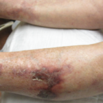
Initially, given the elevated inflammatory markers, new skin lesions and low complements (i.e., C4), the patient was treated with prednisone 60 mg orally once daily for a possible lupus flare. Despite high steroid doses, the patient’s skin lesions continued to spread and worsen. A biopsy result of her skin lesions was obtained, showing diffuse calcium deposits throughout the dermis and subcutaneous layers (including the blood vessels), which was consistent with calciphylaxis.

An extensive workup was performed to determine the etiology of the patient’s calciphylaxis. It involved the evaluation for anti-phospholipid syndrome (APLS), cryoglobulinemia, anti-neutrophil cytoplasmic antibody (ANCA) associated vasculitis, endocarditis, septic emboli, malignancy and hyperparathyroidism. Unfortunately, this workup proved inconclusive. Parathyroid hormone (PTH), parathyroid hormone-related protein (PTHrP), cryoglobulin levels, myeloperoxidase antibodies (MPO), proteinase 3 antibodies (PR3), beta-2-glycoprotein, cardiolipin antibodies, lupus anticoagulant, creatinine kinase and serum protein electrophoresis (SPEP) were all negative or within normal limits.

ad goes here:advert-1
ADVERTISEMENT
SCROLL TO CONTINUE

A computed tomography (CT) scan of the chest, abdomen and pelvis revealed diffuse subcutaneous calcifications and diffuse bilateral groundglass opacities in the lungs (see Figure 2). There were no masses or nodules suspicious for malignancy. A nuclear bone scan demonstrated abnormal radiotracer uptake in bilateral lungs, soft tissues in the upper and lower extremities, and her stomach, which was consistent with calciphylaxis (see Figure 3).

Initially, the patient was started on sodium thiosulfate 25 g IV three times per week. Prednisone was tapered down quickly to 20 mg once a day to aid with wound healing and minimize infection risk. Both mycophenolate mofetil and belimumab were withheld during patient’s initial hospitalization to minimize further infection risk. Despite these efforts, the wounds rapidly worsened over the following week and a half.

ad goes here:advert-2
ADVERTISEMENT
SCROLL TO CONTINUE

Given the concern the patient’s underlying connective tissue disease (CTD) was driving the calciphylaxis, intravenous immunoglobulin therapy (IVIG) was given at a dose of 2 mg/kg split into three consecutive days. However, the patient’s skin lesions continued to spread. Sodium thiosulfate was discontinued after approximately two weeks of therapy, and the patient was given a trial of pamidronate 30 mg IV. Unfortunately, the patient became hypocalcemic and could not receive further pamidronate doses. The patient also received plasmapheresis every other day for a total of five sessions over a two-week period, during which the lesions appeared to stabilize, but then started getting worse again.

The patient was transferred to a nearby hospital for a trial of hyperbaric oxygen therapy, but was deemed not a candidate due to severe mitral regurgitation in the setting of a ruptured chordae tendonae as a result of calciphylaxis affecting the heart structures.
Page: 1 2 3 4 5 6 | Single Page
Share: 

Filed under:Systemic Lupus Erythematosus Tagged with:calciphylaxisconnective tissue diseaseSystemic lupus erythematosus

Related Articles

    Case Report: Warfarin-Induced Non-Uremic Calciphylaxis Mimicking Vasculitis

    May 13, 2021

    Calciphylaxis, or calcific uremic arteriolopathy, is a rare disease characterized by calcification of the arterioles and capillaries in the dermis and subcutaneous tissue, resulting in thrombus formation and subsequent skin ischemia and necrosis.1 This serious condition most commonly occurs in patients with end-stage renal disease (ESRD) requiring dialysis or in kidney transplant recipients. In rare…

    Persistent Symptomatic Hypocalcemia Due to Denosumab: A Case Review

    October 1, 2014

    Patients with osteoporosis and impaired renal function are at risk

    Exploring the Complement System in Human Disease

    February 1, 2010

    Novel disease associations revealed by whole genome screens

    Lost and found

    The History of ACE Inhibitors in Scleroderma Renal Crisis

    February 16, 2021

    Scleroderma renal crisis is a true medical emergency in rheumatology, one that requires prompt diagnosis and treatment. Here, we review the historic introduction of the angiotensin-converting enzyme inhibitors in this context, and highlight management and key questions moving forward. Background Awareness of renal disease in scleroderma dates back many years. The revered physician William Osler…

  • About Us
  • Meet the Editors
  • Issue Archives
  • Contribute
  • Advertise
  • Contact Us
  • Copyright © 2025 by John Wiley & Sons, Inc. All rights reserved, including rights for text and data mining and training of artificial technologies or similar technologies. ISSN 1931-3268 (print). ISSN 1931-3209 (online).
  • DEI Statement
  • Privacy Policy
  • Terms of Use
  • Cookie Preferences