Video: Every Case Tells a Story| Webinar: ACR/CHEST ILD Guidelines in Practice

An official publication of the ACR and the ARP serving rheumatologists and rheumatology professionals

  • Conditions
    • Axial Spondyloarthritis
    • Gout and Crystalline Arthritis
    • Myositis
    • Osteoarthritis and Bone Disorders
    • Pain Syndromes
    • Pediatric Conditions
    • Psoriatic Arthritis
    • Rheumatoid Arthritis
    • Sjögren’s Disease
    • Systemic Lupus Erythematosus
    • Systemic Sclerosis
    • Vasculitis
    • Other Rheumatic Conditions
  • FocusRheum
    • ANCA-Associated Vasculitis
    • Axial Spondyloarthritis
    • Gout
    • Psoriatic Arthritis
    • Rheumatoid Arthritis
    • Systemic Lupus Erythematosus
  • Guidance
    • Clinical Criteria/Guidelines
    • Ethics
    • Legal Updates
    • Legislation & Advocacy
    • Meeting Reports
      • ACR Convergence
      • Other ACR meetings
      • EULAR/Other
    • Research Rheum
  • Drug Updates
    • Analgesics
    • Biologics/DMARDs
  • Practice Support
    • Billing/Coding
    • EMRs
    • Facility
    • Insurance
    • QA/QI
    • Technology
    • Workforce
  • Opinion
    • Patient Perspective
    • Profiles
    • Rheuminations
      • Video
    • Speak Out Rheum
  • Career
    • ACR ExamRheum
    • Awards
    • Career Development
  • ACR
    • ACR Home
    • ACR Convergence
    • ACR Guidelines
    • Journals
      • ACR Open Rheumatology
      • Arthritis & Rheumatology
      • Arthritis Care & Research
    • From the College
    • Events/CME
    • President’s Perspective
  • Search

Case Report: Warfarin-Induced Non-Uremic Calciphylaxis Mimicking Vasculitis

Marie Dardeno, DO, Joshua D. Sparling, MD, & William Monaco, MD  |  Issue: May 2021  |  May 13, 2021

Reticulated violaceous rash of the bilateral lower extremities found on initial presentation.

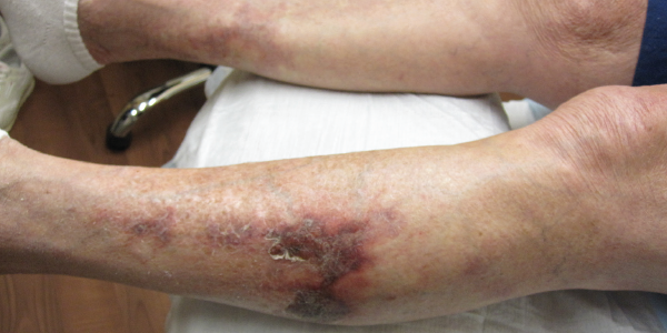
Figure 1. Reticulated violaceous rash of the bilateral lower extremities found on initial presentation.

Approximately six months prior to developing the rash, she was clinically diagnosed with temporal arteritis based on temporal headaches, jaw claudication, scalp tenderness and an excellent response to prednisone. The patient had no history of renal disease, hypothyroidism, liver disease or malignant neoplasm, and no known personal or family history of bleeding or clotting disorders. In addition to warfarin and prednisone, the patient’s medication list at the time of her initial visit also included atorvastatin, quinapril, metoprolol and vitamin D.

On examination, patches of retiform purpura and scattered small (less than 1 cm), necrotic papules on the patient’s lateral calves and medial shins were noted bilaterally. Even though she was taking prednisone, a medium vessel vasculitis with possible occlusive vasculitis was suspected by gross examination on the basis of retiform purpura and the small necrotic ulcerations that had started to develop (see Figure 2).

Development of retiform purpura and the small necrotic ulcerations.

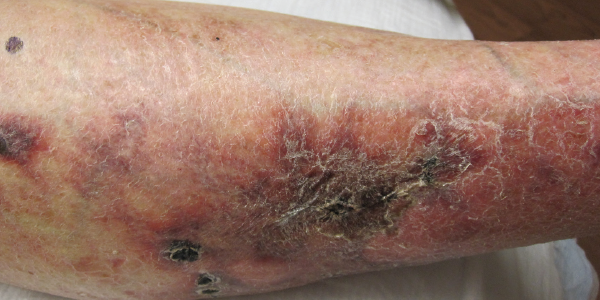
Figure 2. Development of retiform purpura and the small necrotic ulcerations.

Laboratory evaluation yielded negative anti-nuclear antibodies, anti-neutrophil cytoplasmic antibodies, antiphospholipid antibodies, extractable nuclear antigens, rheumatoid factor (<4 IU/mL, normal 0–15 IU/mL), cyclic citrullinated peptic antibody (<0.40 U/mL, normal 0.00–4.99) and serum protein electrophoresis. C3 and C4 complements were both within the normal range, 172.4 mg/dL (normal 90.0–180.0 mg/dL) and 39.1 mg/dL (normal 10.0–40.0 mg/dL), respectively. C-reactive protein was mildly elevated at 3.30 mg/dL (normal 0.00–0.90 mg/dL) and sedimentation rate was within normal range at 10 mm/hr (normal 0–20 mm/hr).

Several skin biopsies were performed, which included an incisional biopsy, a tissue culture biopsy, as well as a biopsy for direct immunofluorescence. Despite these efforts, no definitive histopathological diagnosis, such as vasculitis, calciphylaxis or thrombotic vasculopathy, resulted. In addition, direct immunofluorescence, tissue bacterial cultures and acid-fast cultures were negative. With continued worsening of the patient’s clinical presentation, a drug-induced phenomenon was considered.

ad goes here:advert-2
ADVERTISEMENT
SCROLL TO CONTINUE

Fortunately, the patient was only on a few medications, so warfarin was considered the most likely culprit and was therefore discontinued. Apixaban replaced warfarin for her atrial fibrillation. She was then referred to a tertiary care center for a second opinion. A third set of biopsies finally revealed subcutaneous fibrin thrombi involving the microvasculature, intimal wall thickening and stippled, partially occlusive, calcification consistent with calciphylaxis (see Figures 3 and 4).
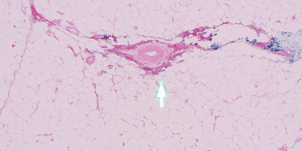
 Figures 3 & 4

Figures 3

H&E and Von Kossa stain, respectively. The third set of biopsies revealed subcutaneous fibrin thrombi involving the microvasculature, intimal wall thickening and stippled, partially occlusive calcification consistent with calciphylaxis.

Figure 4. H&E and Von Kossa stain, respectively. The third set of biopsies revealed subcutaneous fibrin thrombi involving the microvasculature, intimal wall thickening and stippled, partially occlusive calcification consistent with calciphylaxis.

After the diagnosis was made, the patient was started on intravenous infusions of sodium thiosulfate, given the success of this regimen for calcific uremic arteriolopathy.7 The patient returned two months later to the dermatology and rheumatology clinics with significant improvement and minimal resultant scarring from previous necrotic tissue development (see Figure 5, right). Interestingly, the patient reported that improvement seemed to begin soon after discontinuation of the warfarin and prior to beginning sodium thiosulfate.

Page: 1 2 3 4 | Single Page
Share: 

Filed under:ConditionsVasculitis Tagged with:calciphylaxiscase reportEnd-stage renal diseasewarfarin

Related Articles

    Case Report: Refractory Calciphylaxis in Lupus

    May 17, 2018

    Calciphylaxis is a poorly understood and life-threatening ischemic vasculopathy characterized by calcification of the small- and medium-size arteries in the skin, subcutaneous tissue and internal organs, which leads to thrombosis, tissue necrosis and painful skin ulcerations that won’t heal. The disease has a 50–80% mortality rate. Although affected patients typically have end-stage renal disease (ESRD)…

    Case Report: Hydralazine-Induced ANCA-Associated Vasculitis

    February 16, 2021

    Hydralazine has been in use as a treatment for hypertension, most notably in heart failure patients, since 1951.1 The drug is a known cause of autoimmune disease, most specifically hydralazine-induced lupus.  Hydralazine-induced lupus occurs in 7–13% of those taking the medication.2-4 It often presents with constitutional symptoms, arthritis/arthralgias, cutaneous lesions, sero­sitis, myalgias and/or hepatomegaly. Features…

    Exploring the Complement System in Human Disease

    February 1, 2010

    Novel disease associations revealed by whole genome screens

    Effects of Warfarin on Risk of Hip & Knee Replacement in OA Patients

    December 4, 2020

    The use of warfarin may increase the risk of knee or hip replacement in patients with osteoarthritis (OA), according to research presented during ACR Convergence 2020.

  • About Us
  • Meet the Editors
  • Issue Archives
  • Contribute
  • Advertise
  • Contact Us
  • Copyright © 2025 by John Wiley & Sons, Inc. All rights reserved, including rights for text and data mining and training of artificial technologies or similar technologies. ISSN 1931-3268 (print). ISSN 1931-3209 (online).
  • DEI Statement
  • Privacy Policy
  • Terms of Use
  • Cookie Preferences