Although the etiology is unknown, SAM is characterized by four distinctive lesions: mediolysis, separation, arterial gaps and reparative fibrosis.5-8 During the acute of injurious phase, mediolysis results in vacuolization and lysis of the outer arterial media where arterial gaps form, transmural loss of the external elastic lamina occurs and separation of media from the adventitia occurs.9 Aneurysms and dissecting hematomas form as a result, which may lead to sudden massive hemorrhage or vascular occlusion and thrombus formation. Both conditions occurred in our patient.
Acute intra-abdominal or retroperitoneal hemorrhage share the most common causes of increased mortality (20–50%) in the acute phase.10 The reparative phase is characterized by exuberant granulation tissue formation and fibrosis filling arterial gaps. This process remodels the artery and may lead to narrowing and stenosis from granulation tissue overgrowth into the adjacent intact intima. The reparative phase is responsible for the wide variety of angiographic lesions associated with SAM.11
Presentation

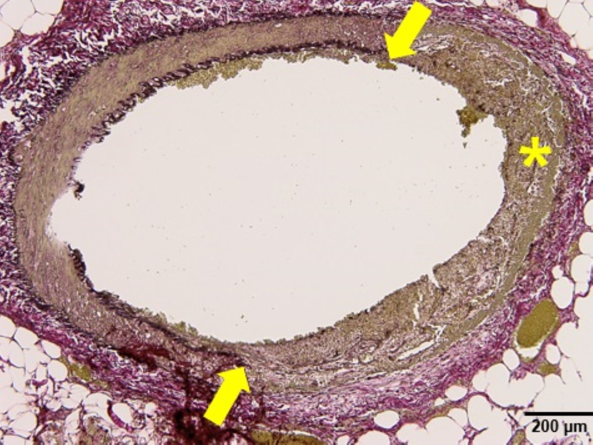
Figure 3b: The EVG special stain (x10 objective) highlights in another artery segmental necrosis of the arterial wall, including absence of both the external and internal elastic lamina, as indicated by the yellow arrows and asterisk. (Click to enlarge.)
Patients with SAM present with symptoms according to the affected vascular bed. The condition most commonly affects the splanchnic vessels, often leading to abdominal apoplexy due to aneurysmal rupture, or symptoms related to ischemia and infarction, as in our case.5,8,12 Less commonly, patients can present with hematuria or acute flank pain when the renal vasculature is involved.7,13–15 Additionally, isolated cases with histopathologic features compatible with SAM have been described in the coronary arteries of neonates, children and young adults, and the cerebral arteries of adults.5–7,16–20
vEDS Introduction
vEDS is a rare autosomal dominant disorder caused by heterogenous mutations in the α1 type III collagen gene COL3A1, leading to a deficit in collagen III.21,22 Collagen III is a major extracellular matrix component of the vasculature and hollow organs. Abnormal or deficient collagen III production leads to arterial, intestinal and uterine fragility, the characteristic features of vEDS.23,24 Different variants at the COL3A1 gene have been shown to influence the course of the disease and clinical phenotype, which contributes to the complexity of making the diagnosis.23 vEDS has the worst prognosis of the Ehlers-Danlos syndromes due to its propensity to rupture hollow organs and arteries. In patients who are symptomatic, arterial complications tend to predominate, followed by digestive and obstetric complications, respectively.23