Video: Every Case Tells a Story| Webinar: ACR/CHEST ILD Guidelines in Practice

An official publication of the ACR and the ARP serving rheumatologists and rheumatology professionals

  • Conditions
    • Axial Spondyloarthritis
    • Gout and Crystalline Arthritis
    • Myositis
    • Osteoarthritis and Bone Disorders
    • Pain Syndromes
    • Pediatric Conditions
    • Psoriatic Arthritis
    • Rheumatoid Arthritis
    • Sjögren’s Disease
    • Systemic Lupus Erythematosus
    • Systemic Sclerosis
    • Vasculitis
    • Other Rheumatic Conditions
  • FocusRheum
    • ANCA-Associated Vasculitis
    • Axial Spondyloarthritis
    • Gout
    • Psoriatic Arthritis
    • Rheumatoid Arthritis
    • Systemic Lupus Erythematosus
  • Guidance
    • Clinical Criteria/Guidelines
    • Ethics
    • Legal Updates
    • Legislation & Advocacy
    • Meeting Reports
      • ACR Convergence
      • Other ACR meetings
      • EULAR/Other
    • Research Rheum
  • Drug Updates
    • Analgesics
    • Biologics/DMARDs
  • Practice Support
    • Billing/Coding
    • EMRs
    • Facility
    • Insurance
    • QA/QI
    • Technology
    • Workforce
  • Opinion
    • Patient Perspective
    • Profiles
    • Rheuminations
      • Video
    • Speak Out Rheum
  • Career
    • ACR ExamRheum
    • Awards
    • Career Development
  • ACR
    • ACR Home
    • ACR Convergence
    • ACR Guidelines
    • Journals
      • ACR Open Rheumatology
      • Arthritis & Rheumatology
      • Arthritis Care & Research
    • From the College
    • Events/CME
    • President’s Perspective
  • Search

Case Review: Lupus Patient with Acute Disseminating Encephalomyelitis

Teresa Sosenko, MD, Anca Musetescu, MD, PhD, Neha Gandhi, MD, Scott Friedstrom, MD, & Diana Girnita, MD, PhD  |  Issue: January 2018  |  January 19, 2018

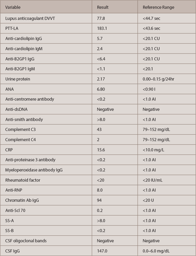
Autoimmune workup revealed positive antinuclear antibody (ANA) (titer 1:640, speckled pattern). ANA subset serology identified positive SS-A, ribonucleoprotein and anti-Smith antibodies. Interestingly, double-stranded DNA (ds-DNA) and SS-B were negative (see Table 2, opposite). An antiphospholipid antibody panel was positive for elevated lupus anticoagulant, but it might have been falsely positive because the patient was already on anticoagulation with a direct thrombin inhibitor started a few weeks before this admission for DVT.

Table 2: The Patient’s Autoimmune Evaluation

(click for larger image)
Table 2: The Patient’s Autoimmune Evaluation

Urinalysis was positive for nephritic range proteinuria (protein in urine was 2.17 g/24 hr); hypoalbuminemia was noted (2.0 g/L) as well. Urine and serum electro­phoresis were also completed, and were negative for monoclonal protein. MRI of the cervical spine and a CT angiogram of the patient’s head and neck were also completed, and showed no signs of vasculitis.

ad goes here:advert-1
ADVERTISEMENT
SCROLL TO CONTINUE

The patient’s clinical situation remained unchanged: He was nonresponsive and had severe paresis on the left side of his body. Because the infectious workup was negative, the team discontinued antibiotics.

Based on these findings, the patient was diagnosed with new-onset SLE with CNS involvement. Both the ACR and the SLICC Criteria for the SLE classification were met, including six ACR criteria and eight SLICC Criteria: serositis, renal disorder, neurologic disorder, hematologic disorder, ANA and immunologic disorder (i.e., ANA, anti-Smith, antiphospholipid and low complement levels).

ad goes here:advert-2
ADVERTISEMENT
SCROLL TO CONTINUE

The patient was started on IV methylprednisolone 1 g daily for five days, followed by IV cyclophosphamide at 0.75 g/m2 body surface. Following IV methyl­prednisolone, oral prednisone 1 mg/kg was continued. The team added hydroxy­chloroquine 400 mg daily shortly after.

Soon after he received this regimen, the patient’s mental status improved dramatically. After one month of physical therapy, he was able to ambulate by himself, and he featured minimal paresis on his left lower extremity. Markers of inflammation decreased to normal, and proteinuria resolved.

After six monthly IV cyclophosphamide infusions, the patient is back to normal, with no residual weakness, and he is ready to resume his job. The oral prednisone will be slowly tapered and discontinued soon. The plan: Continue the monthly therapy with cyclophosphamide up to one year, and then decrease the infusion frequency to every three months.

SLE Discussion

SLE is a severe rheumatic disease. Neuropsychiatric symptoms occur in up to two-thirds of patients and include a wide range of manifestations, including headache, cerebrovascular events, cognitive dysfunction and psychosis, as well as even more serious presentations, such as neuropathies, myelitis and encephalomyelitis.2,3 These symptoms can present alone or concomitantly with other system involvement.3

Page: 1 2 3 4 5 6 | Single Page
Share: 

Filed under:ConditionsMeeting ReportsSystemic Lupus Erythematosus Tagged with:ACR/ARHP Annual MeetingbrainClinicalDiagnosisdruglab testLupusmagnetic resonance imagingMRIoutcomepatient careprednisonepsychological disorderrheumatologistrheumatologySystemic lupus erythematosusTreatmentX-ray

Related Articles
    Atypical Giant Cell Arteritis Case Illustrates Diagnosis, Management Challenges

    Atypical Giant Cell Arteritis Case Illustrates Diagnosis, Management Challenges

    November 14, 2021

    Giant cell arteritis (GCA) is a granulomatous vasculitis of large- and medium-sized arteries, usually affecting the cranial branches of the aortic arch. It is the most common vasculitis, with the highest risk factor being age. Accurate diagnosis and prompt initiation of therapy are of great importance to prevent serious complications, with the most feared being…

    Susac’s Syndrome: Confusion, Expressive Aphasia, Gait Instability

    March 8, 2012

    A 49-year-old man presented to the hospital with confusion, dysarthria, expressive aphasia, and progressive gait instability.

    T Cells in Systemic Lupus Erythematosus

    August 1, 2011

    Progress toward targeted therapy

    Experts Discuss Proposed Giant Cell Arteritis Risk Tool

    April 26, 2018

    A proposed model to predict the risk of giant cell arteritis (GCA) prior to a temporal artery biopsy could help triage patients and guide decision making about the need for biopsy or monitoring (see Figure 1). There’s no specific biomarker for GCA, and GCA can be a “diagnostic conundrum, especially when it presents in an…

  • About Us
  • Meet the Editors
  • Issue Archives
  • Contribute
  • Advertise
  • Contact Us
  • Copyright © 2025 by John Wiley & Sons, Inc. All rights reserved, including rights for text and data mining and training of artificial technologies or similar technologies. ISSN 1931-3268 (print). ISSN 1931-3209 (online).
  • DEI Statement
  • Privacy Policy
  • Terms of Use
  • Cookie Preferences