Video: Every Case Tells a Story| Webinar: ACR/CHEST ILD Guidelines in Practice

An official publication of the ACR and the ARP serving rheumatologists and rheumatology professionals

  • Conditions
    • Axial Spondyloarthritis
    • Gout and Crystalline Arthritis
    • Myositis
    • Osteoarthritis and Bone Disorders
    • Pain Syndromes
    • Pediatric Conditions
    • Psoriatic Arthritis
    • Rheumatoid Arthritis
    • Sjögren’s Disease
    • Systemic Lupus Erythematosus
    • Systemic Sclerosis
    • Vasculitis
    • Other Rheumatic Conditions
  • FocusRheum
    • ANCA-Associated Vasculitis
    • Axial Spondyloarthritis
    • Gout
    • Psoriatic Arthritis
    • Rheumatoid Arthritis
    • Systemic Lupus Erythematosus
  • Guidance
    • Clinical Criteria/Guidelines
    • Ethics
    • Legal Updates
    • Legislation & Advocacy
    • Meeting Reports
      • ACR Convergence
      • Other ACR meetings
      • EULAR/Other
    • Research Rheum
  • Drug Updates
    • Analgesics
    • Biologics/DMARDs
  • Practice Support
    • Billing/Coding
    • EMRs
    • Facility
    • Insurance
    • QA/QI
    • Technology
    • Workforce
  • Opinion
    • Patient Perspective
    • Profiles
    • Rheuminations
      • Video
    • Speak Out Rheum
  • Career
    • ACR ExamRheum
    • Awards
    • Career Development
  • ACR
    • ACR Home
    • ACR Convergence
    • ACR Guidelines
    • Journals
      • ACR Open Rheumatology
      • Arthritis & Rheumatology
      • Arthritis Care & Research
    • From the College
    • Events/CME
    • President’s Perspective
  • Search

Dermatologist, Rheumatologist Discuss Refractory Cutaneous Lupus Case

Richard D. Sontheimer, MD, & Samantha C. Shapiro, MD  |  Issue: September 2022  |  September 5, 2022

As a dermatologist/internist with a career-long subspecialty interest in the cutaneous manifestations of the rheumatic diseases, I found the case of refractory acute cutaneous lupus by Samantha C. Shapiro, MD, in the June 2022 issue of The Rheumatologist intriguing in several ways, and I felt my perspectives on this case might provide additional educational value to the rheumatologist readership.

Diagnosis & Classification

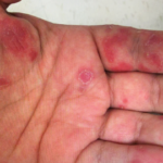
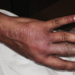
The clinical photos of the patient being discussed suggest a generalized inflammatory skin disorder (i.e., skin lesions both above and below the neck) occurring in the context of a five-year history of systemic lupus erythematosus (SLE). However, the historical duration of the skin changes was not given. The patient’s serologic phenotype was very active at the time of presentation, including anti-double-stranded DNA, RNP, Sm and Ro/SS-A autoantibodies, as well as chronically low serum complement levels. In addition, the patient had leukopenia and thrombocytopenia. However, it is stated that the patient had no internal SLE target-organ disease manifestations.

ad goes here:advert-1
ADVERTISEMENT
SCROLL TO CONTINUE

Had a lesional skin biopsy been performed in this case, it could be presumed it would have demonstrated an interface dermatitis, as would be expected for any form of lupus-specific skin disease. Biopsies of acute cutaneous lupus erythematosus (ACLE) and subacute cutaneous lupus erythematosus (SCLE) lesions have a lymphoid cell-rich inflammatory infiltrate focused at the dermal-epidermal junction with damage to the epidermal basal cell layer (i.e., an interface dermatitis). In addition to displaying an interface dermatitis, biopsies of discoid SLE lesions can also show deep reticular dermis inflammation and damaged skin appendages, such as hair follicles and sweat glands, and result in atrophic scarring. This deeper dermal inflammation in discoid SLE lesions can produce clinical induration, which is characteristically not seen in either ACLE or SCLE lesions.

In the case discussion, it was suggested the skin findings consisted of a combination of ACLE and SCLE lesions. In the early epidemiologic studies of SCLE, it was recognized that SCLE can overlap with either ACLE or classic discoid lupus erythematosus in approximately 20% of cases.1

ad goes here:advert-2
ADVERTISEMENT
SCROLL TO CONTINUE

The case discussion mentioned scarring alopecia on the posterior scalp of this patient. Neither ACLE nor SCLE produce confluent scarring alopecia of the scalp, nor scarring on other parts of the body. In addition, SCLE lesions are characteristically observed on the trunk, with sparing of the central face. These aspects, plus the prominent papulosquamous scale on the skin lesions of this patient, raise the possibility that she could be suffering from a generalized form of classical discoid lupus erythematosus rather than an overlap of ACLE and SCLE. Although uncommon, discoid lupus erythematosus can involve the “butterfly” distribution of facial skin. And generalized classical discoid lupus erythematosus can, at times, be more refractory to treatment than either ACLE or SCLE.

Page: 1 2 3 4 5 6 | Single Page
Share: 

Filed under:ConditionsOpinionSpeak Out RheumSystemic Lupus Erythematosus Tagged with:case reportcutaneous lupus erythematosus (CLE)Dermatologydiagnostic imagingimage caseimage case reportletter to the editor

Related Articles

    Image Case Report: Refractory, Acute, Cutaneous Lupus

    June 14, 2022

    A 25-year-old Mexican American woman with a five-year history of systemic lupus erythematosus (SLE) presents with refractory, acute cutaneous lupus erythematosus (ACLE) and subacute cutaneous lupus erythematosus (SCLE) affecting the scalp, face and hands. Her serologic phenotype is characterized by elevated anti-nuclear, anti-double-stranded deoxyribonucleic acid (dsDNA), anti-ribonucleoprotein (RNP), anti-Smith and anti-SS-A (Ro) antibodies and chronically…

    nukeaf / shutterstock.com

    The History of Treating Lupus with Hydroxychloroquine

    June 15, 2020

    Given how unexpectedly front and center hydroxychloroquine has been in discussions about the treatment of COVID-19 this year, it makes sense to look at how it became so central to the treatment of a rheumatologic condition. In 1991, an article appeared in The New England Journal of Medicine that would alter the way rheumatologists approached…

    Rheumatology Case Report: Bullous Lesions in Patient with Lupus

    April 20, 2017

    Systemic lupus erythematosus (SLE) is a heterogeneous disease associated with multiple acute or chronic cutaneous manifestations, including the relatively rare category of bullous lupus. The development of vesiculo-bullous lesions may be associated with a high morbidity, hence they warrant an urgent investigation, including a skin biopsy to identify the diagnosis and initiate prompt treatment. With…

    2015 ACR/ARHP Annual Meeting: Skin Issues in Rheumatic Diseases Present Challenges

    March 15, 2016

    SAN FRANCISCO—A 40-year-old woman shows up in the clinic with scarring alopecia, with an area of hyperpigmentation on the rim of her scalp, extending from just behind the temple to behind her ears. An examination with a dermatoscope shows hyperkeratotic follicular plugging. ad goes here:advert-1ADVERTISEMENTSCROLL TO CONTINUEThe case—in this example, the discoid form of cutaneous…

  • About Us
  • Meet the Editors
  • Issue Archives
  • Contribute
  • Advertise
  • Contact Us
  • Copyright © 2025 by John Wiley & Sons, Inc. All rights reserved, including rights for text and data mining and training of artificial technologies or similar technologies. ISSN 1931-3268 (print). ISSN 1931-3209 (online).
  • DEI Statement
  • Privacy Policy
  • Terms of Use
  • Cookie Preferences