Video: Every Case Tells a Story| Webinar: ACR/CHEST ILD Guidelines in Practice

An official publication of the ACR and the ARP serving rheumatologists and rheumatology professionals

  • Conditions
    • Axial Spondyloarthritis
    • Gout and Crystalline Arthritis
    • Myositis
    • Osteoarthritis and Bone Disorders
    • Pain Syndromes
    • Pediatric Conditions
    • Psoriatic Arthritis
    • Rheumatoid Arthritis
    • Sjögren’s Disease
    • Systemic Lupus Erythematosus
    • Systemic Sclerosis
    • Vasculitis
    • Other Rheumatic Conditions
  • FocusRheum
    • ANCA-Associated Vasculitis
    • Axial Spondyloarthritis
    • Gout
    • Psoriatic Arthritis
    • Rheumatoid Arthritis
    • Systemic Lupus Erythematosus
  • Guidance
    • Clinical Criteria/Guidelines
    • Ethics
    • Legal Updates
    • Legislation & Advocacy
    • Meeting Reports
      • ACR Convergence
      • Other ACR meetings
      • EULAR/Other
    • Research Rheum
  • Drug Updates
    • Analgesics
    • Biologics/DMARDs
  • Practice Support
    • Billing/Coding
    • EMRs
    • Facility
    • Insurance
    • QA/QI
    • Technology
    • Workforce
  • Opinion
    • Patient Perspective
    • Profiles
    • Rheuminations
      • Video
    • Speak Out Rheum
  • Career
    • ACR ExamRheum
    • Awards
    • Career Development
  • ACR
    • ACR Home
    • ACR Convergence
    • ACR Guidelines
    • Journals
      • ACR Open Rheumatology
      • Arthritis & Rheumatology
      • Arthritis Care & Research
    • From the College
    • Events/CME
    • President’s Perspective
  • Search

Dermatology Case Answer: History of ANCA–Associated Vasculitis, Fever, Rash

Joseph F. Merola, MD, Monica Ramirez, MD  |  Issue: April 2012  |  April 6, 2012

C. Urticaria: The edematous nature of the Sweet’s lesion may, at times, resemble those of robust urticarial lesions; however, there are many clinical features to help distinguish these entities. The lesions of urticaria will migrate over time (excluding the case of urticarial vasculitis) with areas of clearing developing hours after mast cell degranulation has occurred locally. Symptoms such as pruritus further help to differentiate lesions. Systemic symptoms including fever do not occur in common urticaria, although they may be present in urticarial vasculitis and the urticarial eruptions of serum sickness-like reactions, potentially occurring together with arthralgias, malaise, a subtle purpuric component to lesions, and the possible finding of an active urine sediment with renal involvement. Histopathologic evaluation of skin biopsies in these entities would readily distinguish them from Sweet’s syndrome.

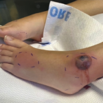
D. Systemic Mycoses/Deep Fungal Infections: Deep fungal infections may present as tender, scattered erythematous nodules and/or plaques. The image of these nodular lesions should invoke a differential diagnosis which includes panniculitides, including the septal panniculitis of the erythema nodosum; small–medium vessel vasculitides involving the skin, such as cutaneous polyarteritis nodosa; subcutaneous Sweet’s syndrome as described above; and infections, particularly deep fungal infections. In the proper clinical context, such as the immunocompromised individual, fungal infection should be considered and therefore a work-up including biopsy for fungal stains and culture should be performed.

ad goes here:advert-1
ADVERTISEMENT
SCROLL TO CONTINUE

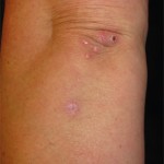
E. Recurrent ANCA-vasculitis with cutaneous involvement: While recurrence of the patient’s disease could certainly present in the skin, her lesions are not typical of a small–medium vessel vasculitis. We would expect the primary lesion in that case to be nonblanching, most classically palpable purpuric lesions of small vessel vasculitis, although some morphologic variation might exist. The biopsy would demonstrate vasculitic changes (fibrinoid necrosis of the vessel wall, leukocytoclasia, extravasated red blood cells, and, in the case of granulomatous polyangiitis, a granulomatous or necrotizing component, as well) if the patient had recurrent disease involving her skin, whereas Sweet’s rarely demonstrates features of vasculitis and would have a much more dramatic neutrophilic infiltrate and papillary dermal edema.

References

  1. Cohen PR. Sweet’s syndrome—a comprehensive review of an acute febrile neutrophilic dermatosis. Orphanet J Rare Dis. 2007;2:34.
  2. Su WPD, Liu HNH. Diagnostic criteria for Sweet’s syndrome. Cutis. 1986;37:167-174.
  3. El-Azhary RA, Brunner KL, Gibson, LE. Sweet’s syndrome as a manifestation of azathioprine hypersensitivity. Mayo Clin Proc. 2008;83:1026-1030.
  4. Paoluzi OA, Crispino P, Amantea A, et al. Diffuse febrile dermatosis in a patient with active ulcerative colitis under treatment with steroids and azathioprine: A case of Sweet’s syndrome. Case report and review of the literature. Dig Liver Dis. 2004;36:361-366.
  5. Yiasemides E, Thom G. Azathioprine hypersensitivity presenting as a neutrophilic dermatosis in a man with ulcerative colitis. Astralas J Dermatol. 2009;50:48-51.
  6. Cohen PR, Kurzrock R. Sweet’s syndrome and cancer. Clin Dermatol. 1993;11:149-157.
  7. Walker DC, Cohen PR. Trimethoprim-sulfamethoxazole-associated acute febrile neutrophilic dermatoses: Case report and review of drug-induced Sweet’s syndrome. J Am Acad Dermatol. 1996;34:918-923.


ad goes here:advert-2
ADVERTISEMENT
SCROLL TO CONTINUE

Page: 1 2 3 | Single Page
Share: 

Filed under:ConditionsVasculitis Tagged with:ANCA-Associated Vasculitismicrobepatient carerheumatologistSteroidsSweet's syndromeVasculitis

Related Articles

    Case Report: Sweet Syndrome as an Initial Presentation of Crohn’s Disease

    July 13, 2022

    Acute febrile neutrophilic dermatosis, or Sweet syndrome, is an inflammatory disease that classically presents with fever, leukocytosis and tender, erythematous plaques characterized by neutrophilic infiltrates on biopsy. Sweet syndrome has been reported in association with several autoimmune diseases, including inflammatory bowel disease, systemic lupus erythematous, rheumatoid arthritis and sarcoidosis.1 Here, we discuss a case of…

    Case Study in Dermatology: Tender Papules on Elbows, Hands in RA

    February 17, 2016

    The Case A 41-year-old woman was referred to the dermatology clinic for a three-month history of tender lesions on her elbows and around the joints of her hands. Her medical history was notable for seropositive, non-erosive rheumatoid arthritis (RA), which was being treated with 5 mg pred­nisone daily, 10 mg methotrexate weekly by mouth, 200…

    Case Report: Hydralazine-Induced ANCA-Associated Vasculitis

    February 16, 2021

    Hydralazine has been in use as a treatment for hypertension, most notably in heart failure patients, since 1951.1 The drug is a known cause of autoimmune disease, most specifically hydralazine-induced lupus.  Hydralazine-induced lupus occurs in 7–13% of those taking the medication.2-4 It often presents with constitutional symptoms, arthritis/arthralgias, cutaneous lesions, sero­sitis, myalgias and/or hepatomegaly. Features…

    Case Report: A 40-Year-Old Man with Vasculitic Neuropathy

    Case Report: A 40-Year-Old Man with Vasculitic Neuropathy

    October 19, 2020

    Ironically, chronic exposure to minocycline has also been associated with a variety of autoimmune syndromes, including drug-induced lupus, auto­immune hepatitis, serum sickness and vasculitis.1 Minocycline is associated with an 8.5-fold increased risk of drug-induced lupus.2 Minocycline and nitrofurantoin are implicated in 90% of cases of drug-induced autoimmune hepatitis.3 Minocycline-induced vasculitis is much less common and,…

  • About Us
  • Meet the Editors
  • Issue Archives
  • Contribute
  • Advertise
  • Contact Us
  • Copyright © 2025 by John Wiley & Sons, Inc. All rights reserved, including rights for text and data mining and training of artificial technologies or similar technologies. ISSN 1931-3268 (print). ISSN 1931-3209 (online).
  • DEI Statement
  • Privacy Policy
  • Terms of Use
  • Cookie Preferences