Video: Every Case Tells a Story| Webinar: ACR/CHEST ILD Guidelines in Practice

An official publication of the ACR and the ARP serving rheumatologists and rheumatology professionals

  • Conditions
    • Axial Spondyloarthritis
    • Gout and Crystalline Arthritis
    • Myositis
    • Osteoarthritis and Bone Disorders
    • Pain Syndromes
    • Pediatric Conditions
    • Psoriatic Arthritis
    • Rheumatoid Arthritis
    • Sjögren’s Disease
    • Systemic Lupus Erythematosus
    • Systemic Sclerosis
    • Vasculitis
    • Other Rheumatic Conditions
  • FocusRheum
    • ANCA-Associated Vasculitis
    • Axial Spondyloarthritis
    • Gout
    • Psoriatic Arthritis
    • Rheumatoid Arthritis
    • Systemic Lupus Erythematosus
  • Guidance
    • Clinical Criteria/Guidelines
    • Ethics
    • Legal Updates
    • Legislation & Advocacy
    • Meeting Reports
      • ACR Convergence
      • Other ACR meetings
      • EULAR/Other
    • Research Rheum
  • Drug Updates
    • Analgesics
    • Biologics/DMARDs
  • Practice Support
    • Billing/Coding
    • EMRs
    • Facility
    • Insurance
    • QA/QI
    • Technology
    • Workforce
  • Opinion
    • Patient Perspective
    • Profiles
    • Rheuminations
      • Video
    • Speak Out Rheum
  • Career
    • ACR ExamRheum
    • Awards
    • Career Development
  • ACR
    • ACR Home
    • ACR Convergence
    • ACR Guidelines
    • Journals
      • ACR Open Rheumatology
      • Arthritis & Rheumatology
      • Arthritis Care & Research
    • From the College
    • Events/CME
    • President’s Perspective
  • Search

Dermatology Case Review

Joseph F. Merola, MD  |  Issue: January 2012  |  January 13, 2012

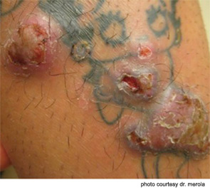
Figure 1: Erythematous, firm nodules with foci of superficial erosion localized to red-containing pigmentary areas of a recently placed tattoo (note involvement of the cartoon’s lips, dress, and heart shape).

Editor’s Note: This month The Rheumatologist is introducing a new feature: Dermatology Case Review. See if you can diagnose the dermatological condition from the photograph and case history.

The Case

A 33-year-old woman presents with somewhat pruritic, nodular lesions worsening over the past three months in the site of a newly placed tattoo. The nodules are restricted to certain portions of the tattoo only (see Figure 1).

ad goes here:advert-1
ADVERTISEMENT
SCROLL TO CONTINUE

What is your diagnosis?

  1. Majocchi’s granuloma
  2. Atypical mycobacterial infection
  3. Hypersensitivity reaction to tattoo particles
  4. Sarcoidosis
  5. Hypertrophic lichen planus

click here for the answer.

Share: 

Filed under:ConditionsOther Rheumatic ConditionsSystemic Sclerosis Tagged with:Dermatologypatient carerheumatologistSarcoidosisScleroderma

Related Articles

    Dermatology Case Answer

    January 13, 2012

    Figure 2: Hematoxylin and eosin stain from punch biopsy of lesional skin demonstrated sarcoidal granulomas, inflamed. Review the case… The correct answer is D) Sarcoidosis: This patient’s first presentation of sarcoidosis consisted of skin lesions that were sarcoidal granulomas localized to a single pigment color within her tattoo (note the red pigment areas only are affected)….

    Case Report: Perplexing Pulmonary Nodules

    March 14, 2022

    Pulmonary nodules are com­mon; most are benign, but the differential diagnosis is broad and includes life-threatening possibilities.1 Our patient is a former smoker who has a history of a complex autoimmune disease and multiple pulmonary nodules. This case was challenging, but clinical, radiographic and histologic clues helped lead to the correct diagnosis. Case Presentation The…

    Case Study in Dermatology: Tender Papules on Elbows, Hands in RA

    February 17, 2016

    The Case A 41-year-old woman was referred to the dermatology clinic for a three-month history of tender lesions on her elbows and around the joints of her hands. Her medical history was notable for seropositive, non-erosive rheumatoid arthritis (RA), which was being treated with 5 mg pred­nisone daily, 10 mg methotrexate weekly by mouth, 200…

    Sonographic Diagnosis of Knuckle Pads

    October 10, 2022

    The Case A 56-year-old white woman was evaluated for a one-year history of painless bumps on the dorsal aspect of the proximal interphalangeal (PIP) joints of both hands and suspected flexor tenosynovitis in her palms. On examination, small cystic nodules without erythema or tenderness were present on the dorsal aspect of several PIP joints (see…

  • About Us
  • Meet the Editors
  • Issue Archives
  • Contribute
  • Advertise
  • Contact Us
  • Copyright © 2025 by John Wiley & Sons, Inc. All rights reserved, including rights for text and data mining and training of artificial technologies or similar technologies. ISSN 1931-3268 (print). ISSN 1931-3209 (online).
  • DEI Statement
  • Privacy Policy
  • Terms of Use
  • Cookie Preferences