The Rheumatologist
COVID-19 NewsACR Convergence
  • Connect with us:
  • Facebook
  • Twitter
  • LinkedIn
  • YouTube
  • Feed
  • Home
  • Conditions
    • Rheumatoid Arthritis
    • SLE (Lupus)
    • Crystal Arthritis
      • Gout Resource Center
    • Spondyloarthritis
    • Osteoarthritis
    • Soft Tissue Pain
    • Scleroderma
    • Vasculitis
    • Systemic Inflammatory Syndromes
    • Guidelines
  • Resource Centers
    • Axial Spondyloarthritis Resource Center
    • Gout Resource Center
    • Psoriatic Arthritis Resource Center
    • Rheumatoid Arthritis Resource Center
    • Systemic Lupus Erythematosus Resource Center
  • Drug Updates
    • Biologics & Biosimilars
    • DMARDs & Immunosuppressives
    • Topical Drugs
    • Analgesics
    • Safety
    • Pharma Co. News
  • Professional Topics
    • Ethics
    • Legal
    • Legislation & Advocacy
    • Career Development
      • Certification
      • Education & Training
    • Awards
    • Profiles
    • President’s Perspective
    • Rheuminations
    • Interprofessional Perspective
  • Practice Management
    • Billing/Coding
    • Quality Assurance/Improvement
    • Workforce
    • Facility
    • Patient Perspective
    • Electronic Health Records
    • Apps
    • Information Technology
    • From the College
    • Multimedia
      • Audio
      • Video
  • Resources
    • Issue Archives
    • ACR Convergence
      • Gout Resource Center
      • Axial Spondyloarthritis Resource Center
      • Psoriatic Arthritis
      • Abstracts
      • Meeting Reports
      • ACR Convergence Home
    • American College of Rheumatology
    • ACR ExamRheum
    • Research Reviews
    • ACR Journals
      • Arthritis & Rheumatology
      • Arthritis Care & Research
      • ACR Open Rheumatology
    • Rheumatology Image Library
    • Treatment Guidelines
    • Rheumatology Research Foundation
    • Events
  • About Us
    • Mission/Vision
    • Meet the Authors
    • Meet the Editors
    • Contribute to The Rheumatologist
    • Subscription
    • Contact
  • Advertise
  • Search
You are here: Home / Articles / Dermatology Case Answer

Dermatology Case Answer

January 13, 2012 • By Joseph F. Merola, MD

  • Tweet
  • Email
Print-Friendly Version / Save PDF
Figure 2: Hematoxylin and eosin stain from punch biopsy of lesional skin demonstrated sarcoidal granulomas, inflamed.

Review the case…

You Might Also Like
  • Dermatology Case Review
  • Dermatology Case Answer: History of ANCA–Associated Vasculitis, Fever, Rash
  • Dermatology Case Answer E: September
Explore This Issue
January 2012
Also By This Author
  • Dermatology Case Review

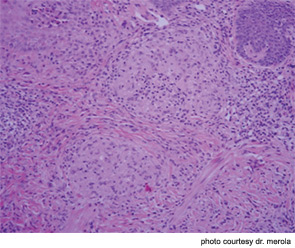
The correct answer is D) Sarcoidosis: This patient’s first presentation of sarcoidosis consisted of skin lesions that were sarcoidal granulomas localized to a single pigment color within her tattoo (note the red pigment areas only are affected). Red tattoo pigment most commonly consists of mercuric sulfide (cinnabar), sienna (ferric hydrate), sandalwood, brazilwood, or organic pigments (aromatic azo compounds). Cutaneous sarcoidosis localized to a tattoo has been well described and several reports demonstrate localization to a single pigment within a given tattoo.

ad goes here:advert-1
ADVERTISEMENT
SCROLL TO CONTINUE

The recommended diagnostic procedure should be a punch biopsy of the affected skin for histopathologic review and culture (see Figure 2). These should include bacterial, fungal, and mycobacterial cultures, since atypical mycobacterial infections from contaminated tattoo materials have been well described. In this case, all tissue cultures were repeatedly negative.

Over time, the chest radiograph demonstrated mediastinal lymphadenopathy consistent with her diagnosis of sarcoid.

ad goes here:advert-2
ADVERTISEMENT
SCROLL TO CONTINUE

Discussion of Other Choices

  1. Majocchi’s granuloma occurs when a superficial fungal/tinea infection extends down into hair follicles and creates a deeper inflammatory granulomatous skin infection. This patient did not have superficial scale or other obvious signs of superficial tinea infection. The biopsy did not demonstrate signs of fungal infection and tissue cultures were negative. Topical antifungal agents may be insufficient to treat these infections.
  2. As discussed above, atypical mycobacterial infection is of particular concern in this patient with a recent tattoo placement. A number of causative mycobacterial strains have been reported, including M. Chelonae, M. Haemophilum, and M. Abscessus.
  3. A granulomatous hypersensitivity reaction may occur, especially to red color–containing pigment (mercury based). This diagnosis was considered until the patient demonstrated other features of sarcoidosis. The possible diagnosis of sarcoidosis should always be considered in these patients.
  4. Hypertrophic lichen planus can present as firm, inflammatory nodules over the lower extremities or more diffusely. They may have a characteristic violaceous appearance. While lichenoid reactions to tattoo pigment have been described, this patient’s pathology results rule out this possibility. Atypical forms of lichen planus, in particular mucosal involvement and hypertrophic variants, may be associated with an underlying hepatitis C infection.

Sources:

  1. Antonovich DD, Callen JP. Development of sarcoidosis in cosmetic tattoos. Arch Dermatol. 2005;141:869-872.
  2. Rodríguez-Blanco I, Fernández LC, Suárez-Peñaranda JM, Pérez del Molino ML, Esteban J, Almagro M. Mycobacterium chelonae infection associated with tattoos. Acta Derm Venereol. 2011;91:61-62.

Dr. Merola

Dr. Merola is an instructor in the Department of Dermatology at Harvard Medical School and a fellow in the Rheumatology Division at Brigham and Women’s Hospital, both in Boston. He is the assistant program director for the Combined Medicine-Dermatology training program and a diplomat of the American Board of Dermatology and the American Board of Internal Medicine.

Pages: 1 2 | Single Page

Filed Under: Conditions, Systemic Inflammatory Syndromes Tagged With: Dermatology, microbe, patient care, radiograph, rheumatologist, SarcoidosisIssue: January 2012

You Might Also Like:
  • Dermatology Case Review
  • Dermatology Case Answer: History of ANCA–Associated Vasculitis, Fever, Rash
  • Dermatology Case Answer E: September
  • Dermatology Case Answer

ACR Convergence

Don’t miss rheumatology’s premier scientific meeting for anyone involved in research or the delivery of rheumatologic care or services.

Visit the ACR Convergence site »

Patient & Caregiver Resources

Find a rheumatology provider. Learn about your condition and how to live with it. English and Spanish language resources.

View Patient & Caregiver Resources »

Meeting Abstracts

Browse and search abstracts from the ACR Convergence and ACR/ARP Annual Meetings going back to 2012.

Visit the Abstracts site »

The Rheumatologist newsmagazine reports on issues and trends in the management and treatment of rheumatic diseases. The Rheumatologist reaches 11,500 rheumatologists, internists, orthopedic surgeons, nurse practitioners, physician assistants, nurses, and other healthcare professionals who practice, research, or teach in the field of rheumatology.

About Us / Contact Us / Advertise / Privacy Policy / Terms of Use / Cookie Preferences

  • Connect with us:
  • Facebook
  • Twitter
  • LinkedIn
  • YouTube
  • Feed

Copyright © 2006–2023 American College of Rheumatology. All rights reserved.

ISSN 1931-3268 (print)
ISSN 1931-3209 (online)