Video: Every Case Tells a Story| Webinar: ACR/CHEST ILD Guidelines in Practice
fa-facebookfa-linkedinfa-youtube-playfa-rss

An official publication of the ACR and the ARP serving rheumatologists and rheumatology professionals

  • Conditions
    • Axial Spondyloarthritis
    • Gout and Crystalline Arthritis
    • Myositis
    • Osteoarthritis and Bone Disorders
    • Pain Syndromes
    • Pediatric Conditions
    • Psoriatic Arthritis
    • Rheumatoid Arthritis
    • Sjögren’s Disease
    • Systemic Lupus Erythematosus
    • Systemic Sclerosis
    • Vasculitis
    • Other Rheumatic Conditions
  • FocusRheum
    • ANCA-Associated Vasculitis
    • Axial Spondyloarthritis
    • Gout
    • Psoriatic Arthritis
    • Rheumatoid Arthritis
    • Systemic Lupus Erythematosus
  • Guidance
    • Clinical Criteria/Guidelines
    • Ethics
    • Legal Updates
    • Legislation & Advocacy
    • Meeting Reports
      • ACR Convergence
      • Other ACR meetings
      • EULAR/Other
    • Research Rheum
  • Drug Updates
    • Analgesics
    • Biologics/DMARDs
  • Practice Support
    • Billing/Coding
    • EMRs
    • Facility
    • Insurance
    • QA/QI
    • Technology
    • Workforce
  • Opinion
    • Patient Perspective
    • Profiles
    • Rheuminations
      • Video
    • Speak Out Rheum
  • Career
    • ACR ExamRheum
    • Awards
    • Career Development
  • ACR
    • ACR Home
    • ACR Convergence
    • ACR Guidelines
    • Journals
      • ACR Open Rheumatology
      • Arthritis & Rheumatology
      • Arthritis Care & Research
    • From the College
    • Events/CME
    • President’s Perspective
  • Search

Envision Arthritis Pathology

Philip G. Conaghan, MB BS, PhD  |  Issue: April 2008  |  April 1, 2008

Until now, RA studies using MRI have been limited to a relatively small number of proof-of-concept or phase II studies. However a number of large randomized RA trials are currently employing MRI, which may eventually result in change of regulatory status.

MRI in RA Clinical Practice

MRI has been responsible for clarifying a number of concepts about RA. First, it has clarified the nature of the relationship between inflammation and damage. It has also helped us understand that subclinical synovitis (i.e., inflammation present in joints that are not detected clinically on joint examination) is common. In a cohort of over 100 patients with low disease activity, over 80% had synovitis when one hand was imaged by MRI.6 The presence of subclinical disease helps to explain the discrepancies when joints that have apparent clinical remission show ongoing radiographic destruction. Such inflammation will make us look harder at what we call “remission” and may lead to us to aim for imaging remission.

ad goes here:advert-1
ADVERTISEMENT
SCROLL TO CONTINUE

Apply caution when jumping from the concepts that MRI has provided to the routine use of MRI in the clinic. At present, there is little information to suggest that MRI is useful in the differential diagnosis for peripheral joints, and defined clinical algorithms are needed before routine use can be advocated. In terms of monitoring erosion progression, there may be a role for the added sensitivity of MRI, especially if it is linked to the better tolerated eMRI. Such use in routine practice would require that individual patient images are always evaluated alongside the previous MR examination, that is, the presence of a systematic longitudinal evaluation whether performed by rheumatologists or radiologists.

FIGURE 2: STIR coronal image of an osteoarthritic knee demonstrating multiple pathologies, especially in the medial compartment where meniscal damage and extrusion, bone marrow edema (femoral condyle), and loss of hyaline cartilage are present.

OA Pathology on MRI

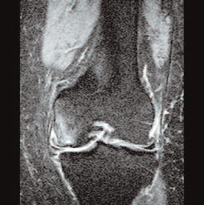
OA is a clinical syndrome for both patients and clinicians. MRI has been a major step forward in understanding the whole organ nature of the structural OA process. However—unlike RA, where MRI has helped simplify our understanding of pathogenesis and treatment response—in OA, we are just beginning to understand the complex interrelationship of pathological processes in multiple tissues. (See Figure 2, right.) MRI has demonstrated OA pathology in the following areas:7

ad goes here:advert-2
ADVERTISEMENT
SCROLL TO CONTINUE

Cartilage: This has long been the focus of much OA research. It has traditionally been evaluated in vivo by using conventional radiographs and measuring the tibio-femoral joint space as a surrogate measure. MRI allows us to examine the cartilage directly, showing that joint space narrowing involves meniscal degeneration and extrusion as well as cartilage loss.

Page: 1 2 3 4 5 6 | Single Page
Share: 

Filed under:ConditionsOsteoarthritis and Bone Disorders Tagged with:Magnetic resonance imaging (MRI)MRIosteoarthritis (OA)PatheogenesisRheumatoid Arthritis (RA)

Related Articles

    Basics of Biologic Joint Reconstruction

    April 6, 2012

    For young patients especially, this can delay knee replacement and provide better outcomes.

    Imaging in Ankylosing Spondylitis

    April 1, 2015

    MRI inflammation, fat and new bone formation in the sacroiliac joints, spine in patients with AS

    Post-Traumatic Osteoarthritis: Managing OA That Develops After Joint Injuries & Reconstructive Surgery

    November 28, 2018

    CHICAGO—Joint trauma is one of many potential drivers of osteoarthritis disease activity and structural progression. In Post-Traumatic OA: Pathogenesis, Clinical Evolution and Management, a session at the 2018 ACR/ARHP Annual Meeting, experts discussed the effects of sports and other injuries on even young patients’ joints. Post-traumatic osteoarthritis (OA) may account for 12% of hip, knee…

    New Therapeutics for Osteoarthritis May Be in Sight

    April 1, 2015

    Overview of OA pathogenesis, recent discoveries suggest new treatment strategies are possible

  • About Us
  • Meet the Editors
  • Issue Archives
  • Contribute
  • Advertise
  • Contact Us
fa-facebookfa-linkedinfa-youtube-playfa-rss
  • Copyright © 2025 by John Wiley & Sons, Inc. All rights reserved, including rights for text and data mining and training of artificial technologies or similar technologies. ISSN 1931-3268 (print). ISSN 1931-3209 (online).
  • DEI Statement
  • Privacy Policy
  • Terms of Use
  • Cookie Preferences