CHICAGO—The lymphatic system took the spotlight in the ACR Convergence 2025 session, Flushing the Joint: Using Lymphatic Drainage to Control Synovial Inflammation, in a basic science talk exploring the role of lymphatics in inflammatory arthritis pathogenesis. The session explored the structure and function of the synovial lymphatic network through a series of elegant experiments, revealing its critical role in modulating synovial inflammation. The talk was delivered by osteoimmunologist Edward Schwarz, PhD, the Burton Professor of Orthopedics and Director of the Center for Musculoskeletal Research at the University of Rochester in Rochester, N.Y.
Introduction to the Lymphatic System
Dr. Schwarz opened the session with a thought-provoking question: If rheumatoid arthritis (RA) is a systemic disease, why is the second metacarpophalangeal (MCP) joint particularly prone to inflammation? As a potential explanation, he projected striking images of a hand injected with fluorescent dye to illustrate significant lymphatic drainage through the second MCP, more so than through the neighboring, smaller MCPs.1
The lymphatic system begins distally as a simple tube of capillaries in the tissues, which function to drain interstitial fluid and inflammatory cells. These capillary tubes converge into afferent collecting lymphatic vessels, which are surrounded by lymphatic muscle cells capable of contraction. These collecting vessels contract to pump fluid to draining lymph nodes, which subsequently return fluid to the venous system.
To illustrate these lymphatic vascular contractions, he displayed images from a mouse model, whereby infrared indocyanine green (first used in oncologic surgery to identify sentinel lymph nodes) was injected into the footpad of a mouse, and lymphatic vessels were seen actively contracting and draining into the more proximal popliteal lymph node.
In acute arthritis mouse models, the contracture frequency of these lymphatic vessels is dramatically increased, while in chronic models, the contractions are lost. Dr. Schwarz encouraged attendees to imagine an acutely inflamed ankle due to rheumatoid arthritis: considering the muscle cell-laden lymphatic vessels reside more proximally up the limb, how do the collecting lymphatic vessels know synovial inflammation is present distally and initiate contractions?
Expanding & Collapsed Lymphatic Phases
In 2007, Proulx et al. demonstrated that in a mouse model of ankle synovitis, the popliteal lymph node was 10–20 times its normal size. They went on to describe the “expanding” and “collapsed” phases of lymph nodes in inflammatory arthritis.2
The expanding phase occurs prior to the onset of distal synovitis, in which there is a large, contrast enhanced, draining lymph node. Eventually, the lymph node will collapse and no longer take up intravenous contrast, which occurs concomitantly with a flare of distal synovitis.3 This expanding and collapsed mechanism also correlates with the velocity of macrophages traveling in the popliteal lymphatic vessels.4
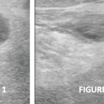
Although first described in mouse models, this phenomenon has since been demonstrated in humans. In acute onset rheumatoid arthritis, magnetic resonance imaging demonstrated expanding popliteal lymph nodes while patients with end-stage, chronic RA have small, collapsed popliteal lymph nodes.5 Similarly, Bell and colleagues demonstrated that healthy hands are able to efficiently drain lymphatic fluid, while patients with active, symptomatic rheumatoid arthritis are have impaired ability to drain indocyanine green, with complete loss of drainage from some lymphatic vessels.6
Lymphatic Models in Inflammatory Arthritis
The lymphatic system is a relatively simple network because there are only two component cells: lymphatic endothelial cells and lymphatic muscle cells. Dr. Schwarz highlighted the work of Scallan et al., who developed an ex vivo system to study the contractile ability of the popliteal lymphatic vessel. Once the lymphatic vessel was removed ex vivo and cannulated, it was flushed with fluid, and they observed coordinated, tonic lymphatic vessel contractions.7
Dr. Schwarz contrasted this coordinated contractile ability with that seen in TNF alpha transgenic mice with inflammatory arthritis—meaning the mice have been genetically engineered to express the TNF-alpha gene leading to inflammation arthritis. In these mice, lymphatic contractions in expanding lymphatics demonstrate an engorged vessel with reduced tonicity and rhythm, while the collapsed lymphatic vessel is essentially non-functional.8 TNF transgenic mice were found to have lymphatic vessels devoid of the normal extracellular matrix, thus reducing contractible ability. Dr. Schwarz then posed the question, what comprises this extracellular matrix and what is its role?
Immersed in the extracellular matrix and buried in the vessel endothelium, popliteal lymphatic vessels are surrounded by mast cells. When these cells are removed or stabilized with cromolyn sodium in a TNF transgenic mouse model, this leads to loss of lymphatic drainage and exacerbation of inflammatory arthritis. This led to the proposal that endothelial mast cells help promote extracellular matrix production and release mediators, such as histamine, to stimulate vessel contractility.9
The Tale of the Telocyte
In an effort to generate a conditional-inducible genetic model in mice for gain and loss of function in lymphatic muscle cells, Dr. Schwarz and his laboratory ultimately discovered the presence of telocytes, which are fibroblastic cells present in all tissues. Telocytes are also known as “nurse cells” given their ability to transmit extracellular matrix, organelles, DNA and RNA into other cells and “nurse” them back to health. Structurally, telocytes have very long dendritic pods calls telopods to communicate with other cell types.
In active synovium of TNF transgenic mice, telocytes are lost, and can be restored with anti-TNF therapy. Electron and confocal microscopy imaging have demonstrated intimate interactions between telocytes and mast cells along lymphatic vessels, leading the team to hypothesize that telocyte signaling to mast cells is essential to histamine release and lymphatic vessel contraction. They further hypothesized and subsequently demonstrated that knocking out telocytes led to a collapsed lymphatic phenotype due to loss of contractile signaling. Telocyte-deficient mice also had a more persistent inflammatory arthritis with greater erosions in a zymosan-induced inflammatory arthritis mouse model.
To summarize the current working model of the synovial lymphatic system and how it drains the joint, Dr. Schwarz said, “there is a telocyte network that extends from telopod to telopod, that parallels the lymphatic vessels from the synovium all the way up to the collecting vessel, where it ends by integrating into mast cells.” Telocytes in the synovium sense osmotic pressure and communicate with mast cells to degranulate and result in a tonic contraction of the vessel.
The model posited two important assumptions, and both were validated experimentally. First, the existence of the network was confirmed via confocal microscopy. Second, investigators demonstrated that telocytes are exquisitely sensitive to osmotic pressure using an in-vitro culture model.
Conclusions
Dr. Schwarz closed with a compelling clinical question: Can exercise maintain or improve telocytes and the synovial lymphatic systemic in chronic inflammatory arthritis? In a mouse model, the answer appears to be yes. He also outlined a working yet intriguing hypothesis that telocytes may differentiate into pro-fibrotic fibroblast like synoviocytes, further contributing to inflammatory arthritis pathogenesis.
During the Q&A, attendees posited that there is likely wider application of this model beyond arthritis, such as photosensitivity in lupus. Dr. Schwarz agreed, adding that a similar process may underlie neural injury in multiple sclerosis.
Although further investigation is needed, the work of Dr. Schwarz and his laboratory team highlighted an important piece of the inflammatory arthritis puzzle—one that may someday be a novel therapeutic target.
Michael Cammarata, MD, RhMSUS, is an assistant professor of medicine at the Johns Hopkins University School of Medicine in Baltimore, Md.
References
- Bouta EM, Bell RD, Rahimi H, et al. Targeting lymphatic function as a novel therapeutic intervention for rheumatoid arthritis. Nat Rev Rheumatol. 2018;14(2):94–106.
- Proulx ST, Kwok E, You Z, et al. Longitudinal assessment of synovial, lymph node, and bone volumes in inflammatory arthritis in mice by in vivo magnetic resonance imaging and microfocal computed tomography. Arthritis Rheum. 2007;56(12):4024–4037.
- Li J, Kuzin I, Moshkani S, et al. Expanded CD23(+)/CD21(hi) B cells in inflamed lymph nodes are associated with the onset of inflammatory-erosive arthritis in TNF-transgenic mice and are targets of anti-CD20 therapy. J Immunol. 2010;184(11):6142–6150.
- Li J, Ju Y, Bouta EM, et al. Efficacy of B cell depletion therapy for murine joint arthritis flare is associated with increased lymphatic flow. Arthritis Rheum. 2013;65(1):130–138.
- Rahimi H, Dieudonne G, Kheyfits V, et al. Relationship Between Lymph Node Volume and Pain Following Certolizumab Therapy for Rheumatoid Arthritis Flare: A Pilot Study. Clin Med Insights Arthritis Musculoskelet Disord. 2016;9:203–208.
- Bell RD, Rahimi H, Kenney HM, et al. Altered Lymphatic Vessel Anatomy and Markedly Diminished Lymph Clearance in Affected Hands of Patients With Active Rheumatoid Arthritis. Arthritis Rheumatol. 2020;72(9):1447–1455.
- Scallan JP, Wolpers JH, Davis MJ. Constriction of isolated collecting lymphatic vessels in response to acute increases in downstream pressure. J Physiol. 2013;591(2):443–459.
- Scallan JP, Bouta EM, Rahimi H, et al. Ex vivoDemonstration of Functional Deficiencies in Popliteal Lymphatic Vessels From TNF-Transgenic Mice With Inflammatory Arthritis. Front Physiol. 2021;12:745096.
- Peng Y, Kenney HM, de Mesy Bentley KL, et al. Distinct mast cell subpopulations within and around lymphatic vessels regulate lymph flow and progression of inflammatory-erosive arthritis in TNF-transgenic mice. Front Immunol. 2023;14:1275871.