Video: Every Case Tells a Story| Webinar: ACR/CHEST ILD Guidelines in Practice

An official publication of the ACR and the ARP serving rheumatologists and rheumatology professionals

  • Conditions
    • Axial Spondyloarthritis
    • Gout and Crystalline Arthritis
    • Myositis
    • Osteoarthritis and Bone Disorders
    • Pain Syndromes
    • Pediatric Conditions
    • Psoriatic Arthritis
    • Rheumatoid Arthritis
    • Sjögren’s Disease
    • Systemic Lupus Erythematosus
    • Systemic Sclerosis
    • Vasculitis
    • Other Rheumatic Conditions
  • FocusRheum
    • ANCA-Associated Vasculitis
    • Axial Spondyloarthritis
    • Gout
    • Psoriatic Arthritis
    • Rheumatoid Arthritis
    • Systemic Lupus Erythematosus
  • Guidance
    • Clinical Criteria/Guidelines
    • Ethics
    • Legal Updates
    • Legislation & Advocacy
    • Meeting Reports
      • ACR Convergence
      • Other ACR meetings
      • EULAR/Other
    • Research Rheum
  • Drug Updates
    • Analgesics
    • Biologics/DMARDs
  • Practice Support
    • Billing/Coding
    • EMRs
    • Facility
    • Insurance
    • QA/QI
    • Technology
    • Workforce
  • Opinion
    • Patient Perspective
    • Profiles
    • Rheuminations
      • Video
    • Speak Out Rheum
  • Career
    • ACR ExamRheum
    • Awards
    • Career Development
  • ACR
    • ACR Home
    • ACR Convergence
    • ACR Guidelines
    • Journals
      • ACR Open Rheumatology
      • Arthritis & Rheumatology
      • Arthritis Care & Research
    • From the College
    • Events/CME
    • President’s Perspective
  • Search

Kussmaul, Meier & Polyarteritis Nodosa

Ruth Jessen Hickman, MD  |  Issue: April 2018  |  April 26, 2018

Seufarth’s symptoms advanced quickly over his 30-day hospitalization, ultimately leading to his death. With the therapeutic tools then available, there was little that Kussmaul could do as treating physician. The authors described Seufarth on the day before his death: “The patient was in a state of extreme weakness. He was scarcely able to speak, lay with persistent severe abdominal and muscle pains, opisthotonically stretched, whimpering, and begged the doctors not to leave him.”1,4

Throughout his illness, the treating clinicians were quite puzzled by the patient’s presentation. Seufarth’s illness could not be fitted into any previously defined clinical picture. “We deemed it wise to forego a diagnosis,” the authors noted. “It was either an unusually complicated trichinosis, or must be an altogether still little known, or unknown illness. It is understandable that under these circumstances, we awaited the results of the autopsy with great anticipation.”1

ad goes here:advert-1
ADVERTISEMENT
SCROLL TO CONTINUE

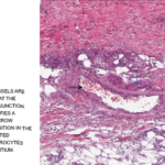
The autopsy report revealed unusual findings of visible nodules along medium-sized arteries. “One glimpse of the transected muscles … immediately demonstrated something unusual … numerous, whitish small tumors up to the size of poppy seeds and hemp seeds.”

Kussmaul and Meier described, “peculiar mostly nodular thickening (periarteritis nodosa) of countless arteries of and below the caliber of the liver artery and the major branches of the coronary arteries of the heart, principally in the bowel, stomach, kidneys, spleen, heart and voluntary muscles, and, to a lesser extent, also in the liver, subcutaneous cell tissue, and in the bronchial and phrenic arteries.” Kussmaul and Meier suggested the term periarteritis nodosa, due to this nodular thickening over blood vessels and the perceived localization of the inflammation to the perivascular sheaths, media and outer layers of the arterial walls.1,4

ad goes here:advert-2
ADVERTISEMENT
SCROLL TO CONTINUE

Now, many other techniques are available to the astute clinicians, including techniques from molecular biology, gene sequencing & detailed medical imaging. Yet when not paired with careful clinical observations, such tests can sometimes lead us astray.

The Paradigmatic Vasculitis

The disease served as a reference point for forms of noninfectious idiopathic vasculitis that were subsequently identified. Its research led to breakthroughs in understanding the pathophysiology of other forms of vasculitis. Dr. Matteson notes, “This description was a really important moment because it became a comparator for other forms of vasculitis that were subsequently described.” Other forms were characterized in terms of their similarities or differences to the constellation of signs, symptoms and pathological findings.5

In the decades to follow, most forms of vasculitis were termed periarteritis nodosa. Even early cases of Kawasaki disease were initially labeled “infantile periarteritis nodosa.” In the early 1900s, the term polyarteritis nodosa began to replace periarteritis nodosa, to emphasize the fact the lesions were found to be transmural and the fact that multiple arteries were affected.4

Page: 1 2 3 4 5 6 | Single Page
Share: 

Filed under:Vasculitis Tagged with:HistoryLost & Foundpolyarteritis nodosascientific methodVasculitis

Related Articles

    Rheum After 5: Dr. Eric Matteson Writes Rheumatic Disease History

    August 12, 2020

    Over the past 20 years, Eric L. Matteson, MD, MPH, emeritus chair, Division of Rheumatology, and emeritus professor of medicine at the Mayo Clinic College of Medicine and Science, Rochester, Minn., as well as a past president of the Rheumatology Research Foundation, has authored or co-authored six books about the history of rheumatic disease and…

    Medium-sized blood vessels are seen in the dermis and at the dermal-subcutaneous junction. The black arrow identifies a venule, and the blue arrow points to a fibrin deposition in the vessel wall. Extravasated neutrophils and erythrocytes are seen in the interstitium.

    Case Report: A Polyarteritis Nodosa Conundrum

    March 14, 2022

    Polyarteritis nodosa (PAN) is a systemic necrotizing vasculitis that typically affects medium-sized muscular arteries. The clinical subsets of PAN are idiopathic, generalized, secondary hepatitis B virus (HBV) associated and cutaneous PAN. These clinical subsets are important because of their therapeutic implications. Virtually any organ system can be affected in generalized PAN, but this vasculitis tends…

    Clues to Diagnosing, Managing Vanishing Aneurysms

    December 16, 2015

    Case report: A 27-year-old male was referred to the rheumatology outpatient department in February 2015 from the urology department after complaining of recent-onset uncontrolled hypertension (220/160 mmHg), headache and vomiting. In 2010, he was admitted to the urologist for sudden-onset left lumbar region pain and recent-onset hypertension. Clinical examination and the blood tests were normal….

    Case Report: Diagnosing, Treating Hepatitis B-Linked Polyarteritis Nodosa

    September 17, 2019

    Hepatitis B virus (HBV) associated polyarteritis nodosa (PAN) is an increasingly rare vasculitis in developed countries due to advances in HBV vaccination and antiviral therapy. However, the condition does persist, and rheumatologists should consider it when evaluating vasculitis cases. Below, we discuss a case that illustrates the varied clinical presentations PAN can encompass. A high…

  • About Us
  • Meet the Editors
  • Issue Archives
  • Contribute
  • Advertise
  • Contact Us
  • Copyright © 2025 by John Wiley & Sons, Inc. All rights reserved, including rights for text and data mining and training of artificial technologies or similar technologies. ISSN 1931-3268 (print). ISSN 1931-3209 (online).
  • DEI Statement
  • Privacy Policy
  • Terms of Use
  • Cookie Preferences