Video: Knock on Wood| Webinar: ACR/CHEST ILD Guidelines in Practice
fa-facebookfa-linkedinfa-youtube-playfa-rss

An official publication of the ACR and the ARP serving rheumatologists and rheumatology professionals

  • Conditions
    • Axial Spondyloarthritis
    • Gout and Crystalline Arthritis
    • Myositis
    • Osteoarthritis and Bone Disorders
    • Pain Syndromes
    • Pediatric Conditions
    • Psoriatic Arthritis
    • Rheumatoid Arthritis
    • Sjögren’s Disease
    • Systemic Lupus Erythematosus
    • Systemic Sclerosis
    • Vasculitis
    • Other Rheumatic Conditions
  • FocusRheum
    • ANCA-Associated Vasculitis
    • Axial Spondyloarthritis
    • Gout
    • Lupus Nephritis
    • Psoriatic Arthritis
    • Rheumatoid Arthritis
    • Systemic Lupus Erythematosus
  • Guidance
    • Clinical Criteria/Guidelines
    • Ethics
    • Legal Updates
    • Legislation & Advocacy
    • Meeting Reports
      • ACR Convergence
      • Other ACR meetings
      • EULAR/Other
    • Research Rheum
  • Drug Updates
    • Analgesics
    • Biologics/DMARDs
  • Practice Support
    • Billing/Coding
    • EMRs
    • Facility
    • Insurance
    • QA/QI
    • Technology
    • Workforce
  • Opinion
    • Patient Perspective
    • Profiles
    • Rheuminations
      • Video
    • Speak Out Rheum
  • Career
    • ACR ExamRheum
    • Awards
    • Career Development
  • ACR
    • ACR Home
    • ACR Convergence
    • ACR Guidelines
    • Journals
      • ACR Open Rheumatology
      • Arthritis & Rheumatology
      • Arthritis Care & Research
    • From the College
    • Events/CME
    • President’s Perspective
  • Search

Latest Research on Cutaneous Vasculitis Diagnosis, Treatment

Lara C. Pullen, PhD  |  Issue: July 2017  |  July 14, 2017

Current guidelines are inadequate to incorporate vasculitic syndromes involving skin.

Current guidelines are inadequate to incorporate vasculitic syndromes involving skin.
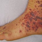
Clinical Photography, Central Manchester University Hospitals NHS Foundation Trust, UK / Science Source

CHICAGO—Warren Piette, MD, professor of dermatology at Rush University Medical Center, Chicago, updated rheumatologists on the topic of cutaneous vasculitis at the ACR’s State-of-the-Art Clinical Symposium in April. He began by explaining that the current vasculitis criteria developed by the ACR in 1990 and the Chapel Hill Consensus Conference (CHCC) in 1994 are inadequate to incorporate vasculitic syndromes involving skin. This deficiency has led an international group to propose revised criteria that will soon be submitted to Arthritis & Rheumatology.

Dr. Piette focused his talk on immune complex vasculitis and anti-neutrophil cytoplasmic autoantibody (ANCA)-associated vasculitis and the purpura that are key to their diagnosis. He explained that purpura can be primary or secondary, and distinguishing the two clinically is more art than science. He emphasized the importance of lesional morphology in sorting purpura pathophysiology into simple hemorrhage, inflammatory hemorrhage or microvascular occlusion, and he also disputed a few dogmas.

ad goes here:advert-1
ADVERTISEMENT
SCROLL TO CONTINUE

Primary Palpable Purpura Are Not Specific for Vasculitis

Primary palpable purpura are not specific for vasculitis. Moreover, explained Dr. Piette, vasculitis is not always palpable or purpuric. He then showed a series of slides of purpura and talked the audience through their diagnoses.

He emphasized the importance of determining if the lesion is blanchable. If the early lesion completely blanches, then it is not purpura, although it could still be vasculitis. If the early lesion partially blanches, then the patient likely has inflammatory purpura, but does not necessarily have vasculitis. Finally, if a lesion doesn’t blanch, then it could simply be a hemorrhage.

ad goes here:advert-2
ADVERTISEMENT
SCROLL TO CONTINUE

Dr. Piette described the four main purpura patterns—multi-generalized, acral, pauci-random and multi-dependent—and showed examples of each. He also noted that there is an evolving recognition that retiform purpura is also a typical morphologic pattern of cutaneous vascular occlusion. Multi-generalized purpura generally contain fewer than 100 lesions, distributed widely and often randomly. They are usually caused by a virus or drug and are only rarely urticarial vasculitis, lymphomatoid papulosis (LyP) and pityriasis lichenoides et varioliformis acuta PLEVA/Lyp, or bowel stasis vasculitis. If the patient with multi-generalized purpura is very ill, however, with multiple lesions retiform, Dr. Piette suggests that purpura fulminans be considered as a diagnosis.

Acral purpura tend to locate on the hand and/or foot. These stocking-glove purpura are likely caused by viral infection, especially parvovirus infection. They may also be caused by sepsis/disseminated intravascular coagulation (DIC) with hypotension leading to microvascular occlusion in vasoconstricted areas. Patients who have retiform purpura in cold-exposed areas, such as ears, nose, hands and feet, likely have cryoglobulins, cryofibrinogens and/or cold agglutinins.

Page: 1 2 3 | Single Page
Share: 

Filed under:ConditionsMeeting ReportsResearch RheumVasculitis Tagged with:2017 State of the Art Clinical SymposiumAC&RAmerican College of Rheumatology (ACR)anti-neutrophilcutaneous vasculitiscytoplasmic autoantibodyDiagnosisGuidelinespatient carepurpuraResearchrheumatologyTreatmentVasculitis

Related Articles

    Rheumatology Case Report: When Moyamoya Disease Mimicks Primary Central Nervous System Vasculitis

    November 16, 2015

    Case report: A 60-year-old Hispanic male with poorly controlled hypertension was sent from the primary care clinic for evaluation of malignant hypertension with a systolic blood pressure above 200 mmHg. His symptoms at the time of presentation included episodic confusion, worsening vision and an unsteady gait. A head computed tomography (CT) scan showed a subacute…

    Case Report: Elderly White Woman Presents with Recurrent Skin Lesions

    Case Report: Elderly White Woman Presents with Recurrent Skin Lesions

    September 17, 2019

    Recurrent cutaneous eosinophilic vasculitis (RCEV) is a rare autoimmune condition characterized histologically by necrotizing small vessel vasculitis of the skin and almost exclusive eosinophilic infiltration without any systemic involvement.1 Frequently, there is associated peripheral eosinophilia, and a prolonged course of glucocorticoids is required for treatment. To date, only a few RCEV cases have been reported….

    Tips for Diagnosing, Treating Seldom Seen Forms of Vasculitis

    February 15, 2017

    WASHINGTON, D.C.—The vast majority of the attention given to vasculitis at the ACR/ARHP Annual Meeting, year after year, focuses on ANCA-associated vasculitis and large-vessel vasculitis, said Philip Seo, MD, MHS, director of the Johns Hopkins Vasculitis Center and moderator of the 2016 ACR Review Course titled, Neglected Vasculitis. That leaves out a lot. “These are…

    Cryopyrin-Associated Periodic Syndromes: Difficult to Recognize, Diagnose, Treat

    October 1, 2014

    Two case studies demonstrate the difficulty, delay in recognizing this rare autoinflammatory disease

  • About Us
  • Meet the Editors
  • Issue Archives
  • Contribute
  • Advertise
  • Contact Us
fa-facebookfa-linkedinfa-youtube-playfa-rss
  • Copyright © 2025 by John Wiley & Sons, Inc. All rights reserved, including rights for text and data mining and training of artificial technologies or similar technologies. ISSN 1931-3268 (print). ISSN 1931-3209 (online).
  • DEI Statement
  • Privacy Policy
  • Terms of Use
  • Cookie Preferences