CHICAGO—Each year at ACR Convergence, the Review Course sessions provide timely and relevant information with speakers who are skilled at distilling complex topics down to their essence. The talks of the first four speakers at this year’s Review Course, on pediatric and adult systemic lupus erythematosus (SLE), inflammatory brain diseases, drug management and mimics of inflammatory myopathies, are summarized here. The talks of the final four speakers are presented below.
Cutaneous Manifestations of Rheumatic Diseases
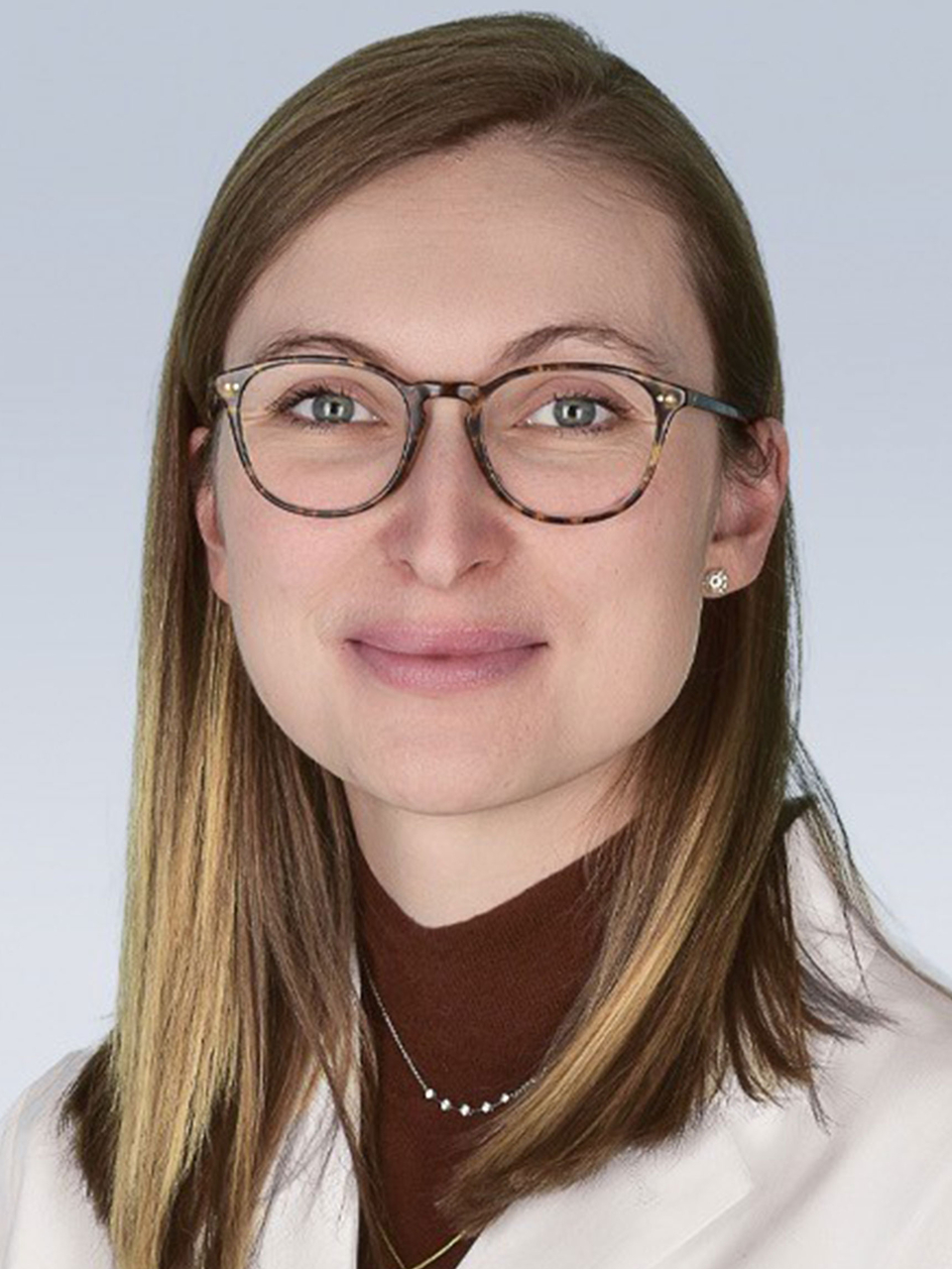
Katharina Shaw, MD, FAAD, director of rheumatology-dermatology at Children’s Hospital of Philadelphia, discussed the cutaneous manifestations of rheumatic diseases. Using a case-based approach, Dr. Shaw very skillfully illustrated that, even in this era of artificial intelligence, a detailed physical exam is often still key to making the correct diagnosis. She provided images of a patient with eosinophilic fasciitis, a condition that is often mistaken for systemic sclerosis, and pointed out specific findings, such as the groove sign, which refers to linear depressions that appear along the course of superficial veins in the affected area, and a cinching of the skin at the waist that she called the corset sign. These help to identify the condition.
Dr. Shaw noted that textbooks indicate the prayer sign, in which inflammatory involvement of the fascia leads to reduced joint mobility and results in an inability to fully extend the fingers at the joints, can be seen in either eosinophilic fasciitis or systemic sclerosis, but clinicians must examine the distal fingers to evaluate for skin laxity, which will be normal in eosinophilic fasciitis and abnormal in systemic sclerosis. As for treatment, pulse therapy with glucocorticoids can be given in severe cases of eosinophilic fasciitis whereas glucocorticoids are generally avoided in patients with systemic sclerosis given the risk of scleroderma renal crisis. Glucocorticoid-sparing treatments for eosinophilic fasciitis can include mycophenolate mofetil, methotrexate, intravenous immunoglobulin and mepolizumab, and patients also benefit from physical therapy. Dr. Shaw noted that it is important to take photos of patients with eosinophilic fasciitis to more objectively evaluate their improvement with treatment, which occurs very slowly.
For lupus, Dr. Shaw reviewed the categories of cutaneous involvement, namely acute, subacute and chronic cutaneous lupus. One form of chronic cutaneous lupus is lupus panniculitis, a condition that most often involves areas of the body with abundant subcutaneous fat, such as the cheeks and temples, upper arms, shoulders, thighs, buttocks and breasts. Indeed, breast involvement—sometimes referred to as lupus mastitis—can mimic breast cancer and may lead to unnecessary diagnostic testing and delay in diagnosis and treatment. Dr. Shaw also told the audience about subcutaneous panniculitis-like T cell lymphoma, a rare, indolent cutaneous lymphoma that typically presents as painless erythematous nodules or plaques, most often on the extremities or trunk. The disease can often be seen in the context of SLE, and diagnosis requires deep skin biopsy and immunophenotyping. Most patients respond well to immunosuppressive therapy, such as glucocorticoids, cyclosporine and methotrexate, with a five-year survival rate exceeding 80%, although patients whose disease is complicated by hemophagocytic lymphohistiocytosis (HLH) have a five-year survival rate of around 46%.1

