Video: Every Case Tells a Story| Webinar: ACR/CHEST ILD Guidelines in Practice

An official publication of the ACR and the ARP serving rheumatologists and rheumatology professionals

  • Conditions
    • Axial Spondyloarthritis
    • Gout and Crystalline Arthritis
    • Myositis
    • Osteoarthritis and Bone Disorders
    • Pain Syndromes
    • Pediatric Conditions
    • Psoriatic Arthritis
    • Rheumatoid Arthritis
    • Sjögren’s Disease
    • Systemic Lupus Erythematosus
    • Systemic Sclerosis
    • Vasculitis
    • Other Rheumatic Conditions
  • FocusRheum
    • ANCA-Associated Vasculitis
    • Axial Spondyloarthritis
    • Gout
    • Psoriatic Arthritis
    • Rheumatoid Arthritis
    • Systemic Lupus Erythematosus
  • Guidance
    • Clinical Criteria/Guidelines
    • Ethics
    • Legal Updates
    • Legislation & Advocacy
    • Meeting Reports
      • ACR Convergence
      • Other ACR meetings
      • EULAR/Other
    • Research Rheum
  • Drug Updates
    • Analgesics
    • Biologics/DMARDs
  • Practice Support
    • Billing/Coding
    • EMRs
    • Facility
    • Insurance
    • QA/QI
    • Technology
    • Workforce
  • Opinion
    • Patient Perspective
    • Profiles
    • Rheuminations
      • Video
    • Speak Out Rheum
  • Career
    • ACR ExamRheum
    • Awards
    • Career Development
  • ACR
    • ACR Home
    • ACR Convergence
    • ACR Guidelines
    • Journals
      • ACR Open Rheumatology
      • Arthritis & Rheumatology
      • Arthritis Care & Research
    • From the College
    • Events/CME
    • President’s Perspective
  • Search

Lupus in the Child’s Mind

Hermine Brunner, MD, MSc, and Marisa Klein-Gitelman, MD, MPH  |  Issue: March 2009  |  March 1, 2009

A pediatric version of the ANAM (Ped-ANAM) software has been designed for use in children who are age 10 years or older; this may be a good screening tool for neurocognitive dysfunction in children with SLE.11 Ped-ANAM testing takes about 30–40 minutes and can be done by the child with little guidance. This test is designed to assess sustained concentration and attention, mental flexibility, spatial processing, cognitive-processing efficiency, arousal/fatigue level, learning, recall, and working memory. More details can be obtained at www.c-shop.ou.edu.

Imaging to Diagnose Neurocognitive Dysfunction

Neuroimaging is a logical means to noninvasively investigate SLE-associated neurocognitive dysfunction, because clinical and laboratory tests have not facilitated timely diagnosis or the understanding of its pathology. Although there has been evidence of cortical and subcortical neuropathology demonstrated by magnetic resonance imaging (MRI), magnetization transfer imaging (MTI), and FLAIR images, correlation of these findings with clinical symptoms has not helped diagnose neurocognitive dysfunction or assess its response to treatment. Findings on conventional MRI have been inconclusive, as lesions are common with SLE in general, and not specifically correlated with, nor sensitive to, change of active neurocognitive dysfunction.

ad goes here:advert-1
ADVERTISEMENT
SCROLL TO CONTINUE

Given the lack of utility of these standard MRI techniques, functional neuroimaging using positron emission tomography and single-photon emission computed tomography have been investigated. Disadvantages of these methods include exposure to radiation and contrast, poor spatial resolution, and a lack of correlation to specific NPSLE syndromes. Metabolic brain studies using magnetic resonance spectrometry (MRS) suggest that the level of cognitive functioning, and NPSLE in general, are associated with both white matter and gray matter changes.12

Figure 2: fMRI images for assessing the effects of childhood-onset SLE on brain activation patterns when testing attention using a continuous performance paradigm. Differences in activation (yellow) and suppression (blue) patterns in children with SLE and healthy controls. In SLE, larger brain areas need to be activated for performing an fMRI task and fewer are suppressed.

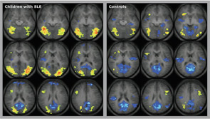
Other recently used techniques include diffusion-weighted imaging (DWI), which can provide quantitative measurements of brain diffusivity.13,14 Interestingly, DWI shows that patients with SLE who test positive for anti-NR2 antibodies have more severe damage to the amygdala compared with SLE patients without anti-NR2 antibodies. The results from advanced imaging techniques, such as diffusion tensor imaging, functional MRI, and diffusion-weighted imaging, in a group of adult patients with various NPSLE syndromes, suggest that the combination of various imaging techniques provides complementary and synergistic information. However, none of these studies is diagnostic.

ad goes here:advert-2
ADVERTISEMENT
SCROLL TO CONTINUE

Page: 1 2 3 4 5 6 7 | Single Page
Share: 

Filed under:ConditionsSystemic Lupus Erythematosus Tagged with:DiagnosisNeurologySystemic lupus erythematosusTreatment

Related Articles

    Seizures in Lupus

    March 1, 2015

    Case report highlights important twist to differential diagnosis of neuropsychiatric lupus

    Pinpoint Cognitive Dysfunction in Patients with Lupus

    April 6, 2012

    More than 80% of SLE patients experience some type of neurologic manifestation during their disease course. The challenge for rheumatologists and other clinicians lies in appropriately diagnosing any cognitive dysfunctions that accompany lupus and better understanding the causes and risk factors of those dysfunctions. “Cognitive Function in SLE” was the focus of a talk at the 2011 ACR/ARHP Annual Scientific Meeting in November.

    Multimodal Imaging May Uncover Neural Mechanisms in Pediatric Neuropsychiatric Lupus

    December 1, 2017

    In clinical practice, many unknowns exist about how SLE affects the brain—particularly in children. To help these young children battling pediatric SLE, Andrea Knight, MD, MSCE, is examining how advanced neuroimaging techniques may uncover potential inflammatory and non-inflammatory mechanisms of neuropsychiatric dysfunction in lupus…

    The Brain in Lupus

    September 1, 2008

    The Mary Kirkland Center lupus conference offers insight into cognitive aspects of SLE

  • About Us
  • Meet the Editors
  • Issue Archives
  • Contribute
  • Advertise
  • Contact Us
  • Copyright © 2025 by John Wiley & Sons, Inc. All rights reserved, including rights for text and data mining and training of artificial technologies or similar technologies. ISSN 1931-3268 (print). ISSN 1931-3209 (online).
  • DEI Statement
  • Privacy Policy
  • Terms of Use
  • Cookie Preferences