Video: Every Case Tells a Story| Webinar: ACR/CHEST ILD Guidelines in Practice

An official publication of the ACR and the ARP serving rheumatologists and rheumatology professionals

  • Conditions
    • Axial Spondyloarthritis
    • Gout and Crystalline Arthritis
    • Myositis
    • Osteoarthritis and Bone Disorders
    • Pain Syndromes
    • Pediatric Conditions
    • Psoriatic Arthritis
    • Rheumatoid Arthritis
    • Sjögren’s Disease
    • Systemic Lupus Erythematosus
    • Systemic Sclerosis
    • Vasculitis
    • Other Rheumatic Conditions
  • FocusRheum
    • ANCA-Associated Vasculitis
    • Axial Spondyloarthritis
    • Gout
    • Psoriatic Arthritis
    • Rheumatoid Arthritis
    • Systemic Lupus Erythematosus
  • Guidance
    • Clinical Criteria/Guidelines
    • Ethics
    • Legal Updates
    • Legislation & Advocacy
    • Meeting Reports
      • ACR Convergence
      • Other ACR meetings
      • EULAR/Other
    • Research Rheum
  • Drug Updates
    • Analgesics
    • Biologics/DMARDs
  • Practice Support
    • Billing/Coding
    • EMRs
    • Facility
    • Insurance
    • QA/QI
    • Technology
    • Workforce
  • Opinion
    • Patient Perspective
    • Profiles
    • Rheuminations
      • Video
    • Speak Out Rheum
  • Career
    • ACR ExamRheum
    • Awards
    • Career Development
  • ACR
    • ACR Home
    • ACR Convergence
    • ACR Guidelines
    • Journals
      • ACR Open Rheumatology
      • Arthritis & Rheumatology
      • Arthritis Care & Research
    • From the College
    • Events/CME
    • President’s Perspective
  • Search

Macrophage Activation Syndrome

Alexei A. Grom, MD  |  Issue: December 2010  |  December 1, 2010

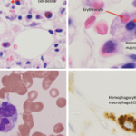
The hemophagocytic macrophages in MAS express CD163.16,17 This feature provides clues to the understanding of the origin of extreme hyperferritinemia in MAS. The only proven function of CD163 is related to its ability to bind hemoglobin– haptoglobin complexes and initiate pathways important for the adaptation to oxidative stress induced by free heme and iron (see Figure 2, p 29).18 In these pathways, to prevent cell damage caused by iron-derived reactive oxygen species, free hemoglobin, the main source of redox-active iron, forms a complex with haptoglobin. The haptoglobin–hemoglobin (Hp–Hb) complexes then bind to CD163 and are internalized by the macrophage. Endocytosis of Hp–Hb complexes leads to upregulation of heme oxygenase (HO) enzymatic activity. HO degrades the heme subunit of Hb into biliverdin, which is subsequently converted to bilirubin, carbon monoxide and free iron. The free iron is either sequestered in association with ferritin within the cell or transported and distributed to red blood cell precursors in the bone marrow. Increased uptake of Hp–Hb complexes by macrophages leads to increased synthesis of ferritin. In MAS, increased release of free Hb associated with erythrophagocytosis would require increased production of ferritin to sequestrate an excessive amount of free iron. Indeed, a strikingly high level of serum ferritin (often above 10,000 ng/dL) is an important diagnostic feature of both MAS and HLH.

Figure 1: Activated macrophages phagocytosing hematopoietic elements in the bone marrow of a systemic JIA patient with MAS.
Source: Prahalad S, Bove KE, Dickens D, Lovell DJ, Grom AA. Etanercept in the treatment of macrophage activation syndrome. J Rheumatol. 2001;28:2120-2124.
Figure 1: Activated macrophages phagocytosing hematopoietic elements in the bone marrow of a systemic JIA patient with MAS. Bone marrow aspirate specimen revealing activated macrophages. (A) Myelocyte within activated macrophage. In addition, there are multiple adherent red blood cell and myeloid precursors. (B) Activated macrophage engulfing a neytrophilic band form. (C) Neutrophilic band forms and metamyelocyte within an activated macrophage. Nuclei of band forms appear condensed a result of destruction. (D) Activated macrophage with hemosiderin deposits and a degenerating phagocytosed nucleated cell.

Clinical Features

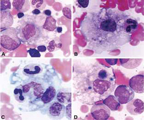
The clinical findings in overt MAS are often dramatic and evolve rapidly. Typically, patients with a chronic condition become acutely ill with persistent fever, mental status changes, lymphadenopathy, hepatosplenomegaly, and liver dysfunction. A hemorrhagic syndrome resembling disseminated intravascular coagulopathy is another striking feature of MAS. Hemorrhagic skin rashes from mild petechiae to extensive ecchymotic lesions, epistaxis, hematemesis secondary to upper gastrointestinal bleeding, and rectal bleeding, are commonly seen in these patients. These clinical symptoms are associated with a precipitous fall in at least two of three blood cell lines (leukocytes, erythrocytes, and platelets). The fall in platelet count is usually an early finding. Because bone marrow aspiration typically reveals significant hypercellularity and normal megakaryocytes, such cytopenias do not seem to be secondary to inadequate production of cells. Increased destruction of the cells by phagocytosis and consumption at the inflammatory sites are more likely explanations.

ad goes here:advert-1
ADVERTISEMENT
SCROLL TO CONTINUE

Page: 1 2 3 4 5 6 7 | Single Page
Share: 

Filed under:Clinical Criteria/GuidelinesConditions Tagged with:Diagnostic CriteriaGuidelinesmacrophage activation syndromeMASPathogenesisTreatment

Related Articles
    Fellows' Forum Case Report: Hemophagocytic Lymphohistiocytosis

    Fellows’ Forum Case Report: Hemophagocytic Lymphohistiocytosis

    August 13, 2017

    The patient was a 48-year-old woman who saw her primary care physician for a flu-like illness three months prior to admission. Her symptoms initially improved, but recurred one month later; she was treated symptomatically, and again symptoms resolved. Two months later, she presented to an outside facility’s emergency department with fever to 103ºF, with associated…

    Diagnosing & Treating Hemophagocytic Lymphohistiocytosis in Adults

    June 15, 2020

    Primary hemophagocytic lymphohistiocytosis (HLH) is caused by genetic mutations and inherited syndromes; it therefore occurs in the pediatric age group. Secondary HLH, however, is more common in adults and is often triggered by other disease states, such as malignancies, chronic immuno­suppression, infections and autoimmune disease.1,2 Macrophage activation syndrome (MAS) is a subset of secondary HLH…

    Ebola: Hemorrhagic Fever and Macrophage Activation Syndrome

    December 1, 2014

    Rheumatologists focus on similar clinical, laboratory findings to better
    understand pathobiology of Ebola infection

    For MAS, Early Diagnosis Is Key

    November 1, 2010

    Research helps hasten diagnosis and treatment of macrophage activation syndrome

  • About Us
  • Meet the Editors
  • Issue Archives
  • Contribute
  • Advertise
  • Contact Us
  • Copyright © 2025 by John Wiley & Sons, Inc. All rights reserved, including rights for text and data mining and training of artificial technologies or similar technologies. ISSN 1931-3268 (print). ISSN 1931-3209 (online).
  • DEI Statement
  • Privacy Policy
  • Terms of Use
  • Cookie Preferences