Video: Every Case Tells a Story| Webinar: ACR/CHEST ILD Guidelines in Practice

An official publication of the ACR and the ARP serving rheumatologists and rheumatology professionals

  • Conditions
    • Axial Spondyloarthritis
    • Gout and Crystalline Arthritis
    • Myositis
    • Osteoarthritis and Bone Disorders
    • Pain Syndromes
    • Pediatric Conditions
    • Psoriatic Arthritis
    • Rheumatoid Arthritis
    • Sjögren’s Disease
    • Systemic Lupus Erythematosus
    • Systemic Sclerosis
    • Vasculitis
    • Other Rheumatic Conditions
  • FocusRheum
    • ANCA-Associated Vasculitis
    • Axial Spondyloarthritis
    • Gout
    • Psoriatic Arthritis
    • Rheumatoid Arthritis
    • Systemic Lupus Erythematosus
  • Guidance
    • Clinical Criteria/Guidelines
    • Ethics
    • Legal Updates
    • Legislation & Advocacy
    • Meeting Reports
      • ACR Convergence
      • Other ACR meetings
      • EULAR/Other
    • Research Rheum
  • Drug Updates
    • Analgesics
    • Biologics/DMARDs
  • Practice Support
    • Billing/Coding
    • EMRs
    • Facility
    • Insurance
    • QA/QI
    • Technology
    • Workforce
  • Opinion
    • Patient Perspective
    • Profiles
    • Rheuminations
      • Video
    • Speak Out Rheum
  • Career
    • ACR ExamRheum
    • Awards
    • Career Development
  • ACR
    • ACR Home
    • ACR Convergence
    • ACR Guidelines
    • Journals
      • ACR Open Rheumatology
      • Arthritis & Rheumatology
      • Arthritis Care & Research
    • From the College
    • Events/CME
    • President’s Perspective
  • Search

Macrophage Activation Syndrome

Alexei A. Grom, MD  |  Issue: December 2010  |  December 1, 2010

A precipitous fall in erythrocyte sedimentation rate (ESR) despite persistently high C-reactive protein is another characteristic laboratory feature, which probably reflects the degree of hypofibrinogenemia secondary to fibrinogen consumption and liver dysfunction. In fact, liver involvement is common in MAS. Significant hepatomegaly is frequently present. Some patients develop mild jaundice. Liver function tests often reveal high serum transaminases activity and mildly elevated levels of serum bilirubin. Moderate hypoalbuminemia has been reported as well. Serum ammonia levels are typically normal or only mildly elevated.

Encephalopathy is another frequently reported clinical feature of MAS. Mental status changes, seizures, and coma are the most common manifestations of central nervous system disease. Cerebrospinal fluid pleiocytosis with mildly elevated protein has been noted in some studies. Significant deterioration in renal function has been noted in several series and was associated with particularly high mortality in one report.3 Pulmonary infiltrates have been mentioned in several reports, and hemophagocytic macrophages can be found in bronchoalveolar lavage.

ad goes here:advert-1
ADVERTISEMENT
SCROLL TO CONTINUE

Additional laboratory findings in MAS include highly elevated serum levels of triglycerides and ferritin. The elevation of ferritin is particularly marked (above 10,000 ng/mL) in most patients and appears to parallel the degree of macrophages activation.

Diagnosis

There are no validated diagnostic criteria for MAS, and early diagnosis is often difficult. Thus, in patients with persistently active underlying rheumatologic disease, a fall in the ESR and platelet count, particularly in a combination with increase in serum D-dimer and ferritin levels, should raise a suspicion of impending MAS. The diagnosis of MAS is usually confirmed by the demonstration of hemophagocytosis in the bone marrow. However, the demonstration of hemophagocytosis in the bone marrow may be difficult due to sampling errors, particularly at the early stages of the syndrome. In addition, it is now increasingly recognized that hemophagocytic macrophages may infiltrate tissues other than bone marrow (e.g., liver, lymph nodes, skin, and lungs). In contrast to MAS, the diagnosis of HLH is usually established based on the validated diagnostic criteria developed by the International Histiocyte Society.9 The criteria include either:

ad goes here:advert-2
ADVERTISEMENT
SCROLL TO CONTINUE
  1. A molecular diagnosis based on specific mutations found in either PRF1 or MUNC13-4 genes; or
  2. A clinical diagnosis based on the following clinical criteria:
      1. Persistent fever;
      2. Splenomegaly;
      3. Cytopenias involving at least two cell lines;
      4. Hypertrigliceremia and/or hypofibrinogenemia;
      5. Hyperferritinemia; and
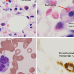
      6. Hemophagocytosis in the bone marrow.

    More recently, two additional laboratory criteria have been added:

    1. Low/absent NK cell cytolytic activity; and
    2. High sIL2Rα levels.

The definite clinical diagnosis of HLH requires the presence of at least five of the eight criteria.

Figure 2: Role of the hemoglobin–haptoglobin scavenger receptor CD163, heme oxygenase, and ferritin in adaptation to oxidative stress induced by free heme and iron. Free heme is a source of redox-active iron.
click for large versionSource: Fall N, Barnes M, Thornton S, et al. Gene expression profiling in peripheral blood in untreated new onset systemic juvenile idiopathic arthritis reveals molecular heterogeneity that may predict macrophage activation syndrome. Arthritis Rheum. 2007;56:3793-3804.

Page: 1 2 3 4 5 6 7 | Single Page
Share: 

Filed under:Clinical Criteria/GuidelinesConditions Tagged with:Diagnostic CriteriaGuidelinesmacrophage activation syndromeMASPathogenesisTreatment

Related Articles
    Fellows' Forum Case Report: Hemophagocytic Lymphohistiocytosis

    Fellows’ Forum Case Report: Hemophagocytic Lymphohistiocytosis

    August 13, 2017

    The patient was a 48-year-old woman who saw her primary care physician for a flu-like illness three months prior to admission. Her symptoms initially improved, but recurred one month later; she was treated symptomatically, and again symptoms resolved. Two months later, she presented to an outside facility’s emergency department with fever to 103ºF, with associated…

    Diagnosing & Treating Hemophagocytic Lymphohistiocytosis in Adults

    June 15, 2020

    Primary hemophagocytic lymphohistiocytosis (HLH) is caused by genetic mutations and inherited syndromes; it therefore occurs in the pediatric age group. Secondary HLH, however, is more common in adults and is often triggered by other disease states, such as malignancies, chronic immuno­suppression, infections and autoimmune disease.1,2 Macrophage activation syndrome (MAS) is a subset of secondary HLH…

    Ebola: Hemorrhagic Fever and Macrophage Activation Syndrome

    December 1, 2014

    Rheumatologists focus on similar clinical, laboratory findings to better
    understand pathobiology of Ebola infection

    For MAS, Early Diagnosis Is Key

    November 1, 2010

    Research helps hasten diagnosis and treatment of macrophage activation syndrome

  • About Us
  • Meet the Editors
  • Issue Archives
  • Contribute
  • Advertise
  • Contact Us
  • Copyright © 2025 by John Wiley & Sons, Inc. All rights reserved, including rights for text and data mining and training of artificial technologies or similar technologies. ISSN 1931-3268 (print). ISSN 1931-3209 (online).
  • DEI Statement
  • Privacy Policy
  • Terms of Use
  • Cookie Preferences