Video: Every Case Tells a Story| Webinar: ACR/CHEST ILD Guidelines in Practice

An official publication of the ACR and the ARP serving rheumatologists and rheumatology professionals

  • Conditions
    • Axial Spondyloarthritis
    • Gout and Crystalline Arthritis
    • Myositis
    • Osteoarthritis and Bone Disorders
    • Pain Syndromes
    • Pediatric Conditions
    • Psoriatic Arthritis
    • Rheumatoid Arthritis
    • Sjögren’s Disease
    • Systemic Lupus Erythematosus
    • Systemic Sclerosis
    • Vasculitis
    • Other Rheumatic Conditions
  • FocusRheum
    • ANCA-Associated Vasculitis
    • Axial Spondyloarthritis
    • Gout
    • Psoriatic Arthritis
    • Rheumatoid Arthritis
    • Systemic Lupus Erythematosus
  • Guidance
    • Clinical Criteria/Guidelines
    • Ethics
    • Legal Updates
    • Legislation & Advocacy
    • Meeting Reports
      • ACR Convergence
      • Other ACR meetings
      • EULAR/Other
    • Research Rheum
  • Drug Updates
    • Analgesics
    • Biologics/DMARDs
  • Practice Support
    • Billing/Coding
    • EMRs
    • Facility
    • Insurance
    • QA/QI
    • Technology
    • Workforce
  • Opinion
    • Patient Perspective
    • Profiles
    • Rheuminations
      • Video
    • Speak Out Rheum
  • Career
    • ACR ExamRheum
    • Awards
    • Career Development
  • ACR
    • ACR Home
    • ACR Convergence
    • ACR Guidelines
    • Journals
      • ACR Open Rheumatology
      • Arthritis & Rheumatology
      • Arthritis Care & Research
    • From the College
    • Events/CME
    • President’s Perspective
  • Search

Melioidosis: What Rheumatologists Need to Know

G.C. Yathish, MD, Taral Parikh, MD, Parikshit Sagdeo, MD, Balakrishnan Canchi, MD, and Gurmeet Mangat, MD  |  Issue: May 2015  |  May 15, 2015

Burkholderia pseudomallei bacteria on a color-enhanced scanning electron micrograph (SEM).

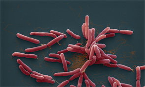
Burkholderia pseudomallei bacteria on a color-enhanced scanning electron micrograph (SEM). Image Credit: Eye of Science / Science Source

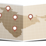
Burkholderia pseudomallei, the causative agent of melioidosis, is endemic in Southeast Asia and northern Australia.1 In recent years, the incidence of melioidosis has increased worldwide. Septic arthritis is a rare, but well-recognized, manifestation of melioidosis.

Case Report

A 49-year-old woman with known diabetes was admitted elsewhere with uncontrolled diabetes and fever. She was found to be in diabetic ketoacidosis. During her hospital stay, she had acute onset of severe pain, swelling and redness over the right shoulder joint. The shoulder joint was surgically drained, empiric parenteral antibiotics were initiated, and the patient was transferred to our hospital for further management.

ad goes here:advert-1
ADVERTISEMENT
SCROLL TO CONTINUE

On examination, she was febrile, the right shoulder was tender and warm, and all range of movement was severely restricted. There was also erythematous swelling of the right second proximal interphalangeal (PIP) joint, and diffuse swelling and tenderness over the left calf area.

Table 1: Salient Features of Melioidosis

(click for larger image)

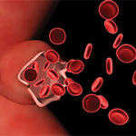
Laboratory results included the following: hemoglobin, 9.8 gm/dL; leukocyte count, 13,400 cu/mm3 (90% neutrophils); platelet count, 4.36 cu/mm3; ESR, 106 mm/hr; CRP, 96 mg/dL; SGPT, 48 IU/L; creatinine, 0.8 mg/dL; rheumatoid factor and anti-CCP antibody were both negative. Lower limb color Doppler ultra­sonography showed a lower left limb intramuscular fluid collection suggestive of an abscess.

ad goes here:advert-2
ADVERTISEMENT
SCROLL TO CONTINUE

An MRI of the right shoulder showed multiple abscesses with osteomyelitic changes (see Figures 1 and 2).

The synovial fluid culture from the shoulder, from the referring hospital, showed gram-negative bacilli on smear, but the organism was not identified. She was switched to injectable cefipime and tazobactam.

She underwent incision and drainage of the right shoulder joint and the left leg abscess. On the third postoperative day, she developed acute-onset tachypnea, and a chest X-ray showed changes of acute respiratory distress syndrome (see Figures 3, 4 and 5). The patient was intubated and treated with parenteral meropenem. However, she became anuric and succumbed to hyperkalemia-induced arrhythmia and cardiac arrest.

The day after her death, bacterial culture reports confirmed the growth of Burkholderia pseudomallei (B. pseudomallei) from the synovial fluid.

MRIs of the right shoulder showing a collection within the right axilla (Fig. 1, top) and hyperintense signals from the head of the right humerus (Fig. 2).

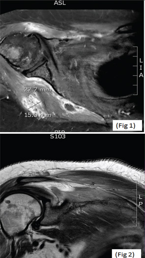
(click for larger image) MRIs of the right shoulder showing a collection within the right axilla (Fig. 1, top) and hyperintense signals from the head of the right humerus (Fig. 2).

Discussion

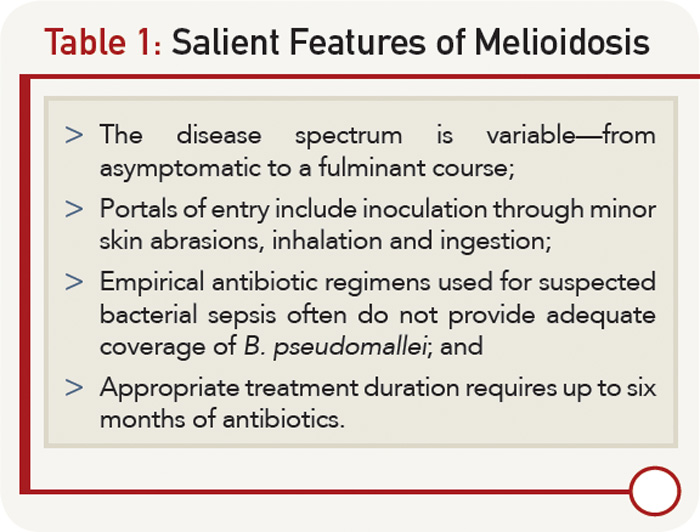
Melioidosis, also known as pseudoglanders or Whitmore’s disease, is caused by a gram-negative bacillus previously classified under genus pseudomonas—B. pseudomallei. It was identified in Burma in 1911 by Whitmore and Krishnaswami. It is classically characterized by the development of pneumonia and multiple abscesses, with a mortality rate as high as 40%. Melioidosis is an important cause of community-acquired sepsis in Southeast Asia and Australia and is classified as a Group B bioterrorism agent.1,2

Page: 1 2 3 | Single Page
Share: 

Filed under:Conditions Tagged with:infectious diseasemelioidosisPainrheumatologistseptic arthritis

Related Articles

    Fellow’s Forum Case Report: Aneurysm in Takayasu’s Arteritis

    August 17, 2015

    A healthy 30-year-old Indian male was performing his routine workout in the gym when he developed giddiness and transient blackouts. He thought it was due to exercising excessively. Over the next month, he developed a low-grade fever and started feeling lethargic, to the extent that he was unable to exercise or work. He was admitted…

    How to Evaluate Shoulder Pain

    February 2, 2013

    The common causes of shoulder pain and diagnostic tests that rheumatologists need to know

    On the Road in Rajasthan: Vehicular-Caused Bone, Joint Damage in India

    March 20, 2017

    In the good old days, physicians routinely made house calls. The decision to visit the literal bedside of a patient was practical: hospital services were primitive and often offered too little benefit to justify an emergency journey by the patient. These physicians carried leather bags, sometimes called Gladstones, that were filled with instruments for eventualities…

    Rheumatologist Fellow Works in India Via ACR Exchange Program

    March 17, 2018

    In November 2017, I went to Lucknow, India, where I would spend my time as an exchange fellow at the Sanjay Gandhi Postgraduate Institute of Medical Sciences (SGPIMS) as part of the ACR International Visiting Fellows Exchange Program. Where I Come From I completed my medical degree at Mahatma Gandhi Missions Medical College, Navi Mumbai…

  • About Us
  • Meet the Editors
  • Issue Archives
  • Contribute
  • Advertise
  • Contact Us
  • Copyright © 2025 by John Wiley & Sons, Inc. All rights reserved, including rights for text and data mining and training of artificial technologies or similar technologies. ISSN 1931-3268 (print). ISSN 1931-3209 (online).
  • DEI Statement
  • Privacy Policy
  • Terms of Use
  • Cookie Preferences