Video: Every Case Tells a Story| Webinar: ACR/CHEST ILD Guidelines in Practice

An official publication of the ACR and the ARP serving rheumatologists and rheumatology professionals

  • Conditions
    • Axial Spondyloarthritis
    • Gout and Crystalline Arthritis
    • Myositis
    • Osteoarthritis and Bone Disorders
    • Pain Syndromes
    • Pediatric Conditions
    • Psoriatic Arthritis
    • Rheumatoid Arthritis
    • Sjögren’s Disease
    • Systemic Lupus Erythematosus
    • Systemic Sclerosis
    • Vasculitis
    • Other Rheumatic Conditions
  • FocusRheum
    • ANCA-Associated Vasculitis
    • Axial Spondyloarthritis
    • Gout
    • Psoriatic Arthritis
    • Rheumatoid Arthritis
    • Systemic Lupus Erythematosus
  • Guidance
    • Clinical Criteria/Guidelines
    • Ethics
    • Legal Updates
    • Legislation & Advocacy
    • Meeting Reports
      • ACR Convergence
      • Other ACR meetings
      • EULAR/Other
    • Research Rheum
  • Drug Updates
    • Analgesics
    • Biologics/DMARDs
  • Practice Support
    • Billing/Coding
    • EMRs
    • Facility
    • Insurance
    • QA/QI
    • Technology
    • Workforce
  • Opinion
    • Patient Perspective
    • Profiles
    • Rheuminations
      • Video
    • Speak Out Rheum
  • Career
    • ACR ExamRheum
    • Awards
    • Career Development
  • ACR
    • ACR Home
    • ACR Convergence
    • ACR Guidelines
    • Journals
      • ACR Open Rheumatology
      • Arthritis & Rheumatology
      • Arthritis Care & Research
    • From the College
    • Events/CME
    • President’s Perspective
  • Search

Musculoskeletal Ultrasound: A Valuable Tool for Diagnosing Rheumatic Illnesses

Khushboo Sheth, MBBS, & Christopher Scola, MD  |  Issue: October 2015  |  October 15, 2015

Image 10: This image was taken after aspiration of the fluid from the cyst.

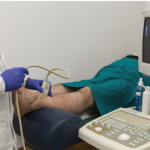
(click for larger image)
Image 10: This image was taken after aspiration of the fluid from the cyst.

Conclusion

MSK ultrasound is an imaging modality with practical application for the clinical rheumatologist. A growing literature supports its reliability in diagnosing early RA and assessing disease activity in established patients in addition to improving accuracy by excluding RA mimics. The added benefit of needle guidance can increase the diagnostic yield of arthrocentesis, which may obviate the need for radiology referral for root joint aspirations (hip and shoulder) or deep bursal aspirations.

Further study of this imaging modality and recognition of its application for the clinician will enhance treatment of rheumatic disease patients.

ad goes here:advert-1
ADVERTISEMENT
SCROLL TO CONTINUE

Khushboo Sheth, MBBS, is a resident in Internal Medicine at the University of Connecticut in Farmington, Conn.

Christopher Scola, MD, is chief of the Rheumatology Division at Hartford Hospital and assistant clinical professor of medicine at the University of Connecticut School of Medicine, Hartford, Conn.

ad goes here:advert-2
ADVERTISEMENT
SCROLL TO CONTINUE

References

  1. American College of Rheumatology. Education and Careers.
  2. Hobson-Webb LD, Massey JM, Juel VC, Sanders DB. The ultrasonographic wrist-to-forearm median nerve area ratio in carpal tunnel syndrome. Clin Neurophysiol. 2008 Jun;119(6):1353–1357.
  3. Aletaha D, Neogi T, Silman AJ, et al. 2010 rheumatoid arthritis classification criteria: An American College of Rheumatology/European League Against Rheumatism collaborative initiative. Arthritis Rheum. 2010 Sep;62(9):2569–2581.
  4. Colebatch AN, Edwards CJ, Ostergaard M, et al. EULAR recommendations for the use of imaging of the joints in the clinical management of rheumatoid arthritis. Ann Rheum Dis. 2013 Jun;72(6):804–814.
  5. Gutierrez M, Schmidt WA, Thiele RG, et al. International consensus for ultrasound lesions in gout: Results of delphi process and Web-reliability exercise. Rheumatology (Oxford). 2015 May 13; pii.
  6. Perez-Ruiz F, Dalbeth N, Urresola A, et al. Imaging of gout: Findings and utility. Arthritis Res Ther. 2009;11(3):232.
  7. Grassi W, Okano T, Filippucci E. Use of ultrasound for diagnosis and monitoring of outcomes in crystal arthropathies. Curr Opin Rheumatol. 2015 Mar;27(2):147–155.
  8. Taylor WJ, Fransen J, Jansen TL, et al. Study for updated gout classification criteria (SUGAR): Identification of features to classify gout. Arthritis Care Res (Hoboken). 2015 Mar 16.
  9. Wakefield RJ, D’Agostino MA. Essential Applications of Musculoskeletal Ultrasound in Rheumatology: Expert Consult, premium ed. Elsevier Health Sciences. 2010.
  10. Frediani B, Filippou G, Falsetti P, et al. Diagnosis of calcium pyrophosphate dihydrate crystal deposition disease: Ultrasonographic criteria proposed. Ann Rheum Dis. 2005 Apr;64(4):638–640.
  11. Zhang W, Doherty M, Bardin T, et al. European League Against Rheumatism recommendations for calcium pyrophosphate deposition. part I: Terminology and diagnosis. Ann Rheum Dis. 2011 Apr;70(4):563–570.

Page: 1 2 3 4 5 6 7 | Single Page
Share: 

Filed under:ConditionsRheumatoid Arthritis Tagged with:ClinicalDiagnosisMSKMusculoskeletal Ultrasoundpatient careRARheumatoid arthritisrheumatologistsrheumatologyTrainingUltrasound

Related Articles

    Ultrasound Can be Useful in Diagnosing Gout

    June 14, 2017

    The presence of synovial monosodium urate monohydrate (MSU) crystals is the gold standard for diagnosing gout. But a new study, funded in part by the ACR and led by rheumatologists, including Alexis Ogdie, MD, MSCE, evaluated the effectiveness of ultrasound in diagnosing it. The study found that ultrasound can be useful in discriminating gout from non-gout….

    Hyperuricemia Doesn’t Always Indicate Gout in Rheumatic Disease

    November 9, 2017

    A 57-year-old Ghanaian woman was referred to our rheumatology practice with acute, left elbow swelling and pain. The referring oncologist suspected gout, because the patient had hyperuricemia. Six months before, the patient was diagnosed with stage IV human T-lymphotropic virus type 1 (HTLV-1)-associated adult T cell lymphoma (ATLL). Her initial oncologic manifestations included multiple thoracic,…

    Sonographic Diagnosis of Knuckle Pads

    October 10, 2022

    The Case A 56-year-old white woman was evaluated for a one-year history of painless bumps on the dorsal aspect of the proximal interphalangeal (PIP) joints of both hands and suspected flexor tenosynovitis in her palms. On examination, small cystic nodules without erythema or tenderness were present on the dorsal aspect of several PIP joints (see…

    The Role Ultrasound Imaging Plays in Diagnosing Hemangiomas

    April 15, 2022

    A 17-year-old woman presents with chronic finger pain experienced over six months that is worse in the mornings. On physical exam, the patient has no joint swelling, pain on range of motion or limitation of range of motion in any of her finger joints. She has a tender, subcutaneous, firm, flesh-colored nodule on the lateral…

  • About Us
  • Meet the Editors
  • Issue Archives
  • Contribute
  • Advertise
  • Contact Us
  • Copyright © 2025 by John Wiley & Sons, Inc. All rights reserved, including rights for text and data mining and training of artificial technologies or similar technologies. ISSN 1931-3268 (print). ISSN 1931-3209 (online).
  • DEI Statement
  • Privacy Policy
  • Terms of Use
  • Cookie Preferences